{"title":"利用数字扫描技术制备的3单元单片氧化锆修复体的边缘和内部配合度的体外评价。","authors":"Çise Özal, Mutahhar Ulusoy","doi":"10.4047/jap.2021.13.6.373","DOIUrl":null,"url":null,"abstract":"<p><strong>Purpose: </strong>This study aimed to compare the marginal and internal fit of 3-unit monolithic zirconia restorations that were designed by using the data obtained with the aid of intraoral and laboratory scanners.</p><p><strong>Materials and methods: </strong>For the fabrication of 3-unit monolithic zirconia restorations using impressions taken from the maxillary master cast, plaster cast was created and scanned in laboratory scanners (InEos X5 and D900L). The main cast was also scanned with different intraoral scanners (Omnicam [OMNI], Primescan [PS], Trios 3 [T3], Trios 4 [T4]) (n = 12 per group). Zirconia fixed partial dentures were virtually designed, produced from presintered block, and subsequently sintered. Marginal and internal discrepancy values (in µm) were measured by using silicone replica method under stereomicroscope. Data were statistically analyzed by using 1-way ANOVA and Kruskal Wallis tests (<i>P</i><.05).</p><p><strong>Results: </strong>In terms of marginal adaptation, the measurements on the canine tooth indicated better performance with intraoral scanners than those in laboratory scanners, but there was no difference among intraoral scanners (<i>P</i><.05). In the premolar tooth, PS had the lowest marginal (86.9 ± 19.2 µm) and axial (92.4 ± 14.8 µm), and T4 had the lowest axio-occlusal (89.4 ± 15.6 µm) and occlusal (89.1 ± 13.9 µm) discrepancy value. In both canine and premolar teeth, the D900L was found to be the most marginally and internally inconsistent scanner.</p><p><strong>Conclusion: </strong>Within the limits of the study, marginal and internal discrepancy values were generally lower in intraoral scanners than in laboratory scanners. Marginal discrepancy values of scanners were clinically acceptable (< 120 µm), except D900L.</p>","PeriodicalId":51291,"journal":{"name":"Journal of Advanced Prosthodontics","volume":"13 6","pages":"373-384"},"PeriodicalIF":2.5000,"publicationDate":"2021-12-01","publicationTypes":"Journal Article","fieldsOfStudy":null,"isOpenAccess":false,"openAccessPdf":"https://ftp.ncbi.nlm.nih.gov/pub/pmc/oa_pdf/4a/dc/jap-13-373.PMC8712113.pdf","citationCount":"4","resultStr":"{\"title\":\"<i>In-vitro</i> evaluation of marginal and internal fit of 3-unit monolithic zirconia restorations fabricated using digital scanning technologies.\",\"authors\":\"Çise Özal, Mutahhar Ulusoy\",\"doi\":\"10.4047/jap.2021.13.6.373\",\"DOIUrl\":null,\"url\":null,\"abstract\":\"<p><strong>Purpose: </strong>This study aimed to compare the marginal and internal fit of 3-unit monolithic zirconia restorations that were designed by using the data obtained with the aid of intraoral and laboratory scanners.</p><p><strong>Materials and methods: </strong>For the fabrication of 3-unit monolithic zirconia restorations using impressions taken from the maxillary master cast, plaster cast was created and scanned in laboratory scanners (InEos X5 and D900L). The main cast was also scanned with different intraoral scanners (Omnicam [OMNI], Primescan [PS], Trios 3 [T3], Trios 4 [T4]) (n = 12 per group). Zirconia fixed partial dentures were virtually designed, produced from presintered block, and subsequently sintered. Marginal and internal discrepancy values (in µm) were measured by using silicone replica method under stereomicroscope. Data were statistically analyzed by using 1-way ANOVA and Kruskal Wallis tests (<i>P</i><.05).</p><p><strong>Results: </strong>In terms of marginal adaptation, the measurements on the canine tooth indicated better performance with intraoral scanners than those in laboratory scanners, but there was no difference among intraoral scanners (<i>P</i><.05). In the premolar tooth, PS had the lowest marginal (86.9 ± 19.2 µm) and axial (92.4 ± 14.8 µm), and T4 had the lowest axio-occlusal (89.4 ± 15.6 µm) and occlusal (89.1 ± 13.9 µm) discrepancy value. In both canine and premolar teeth, the D900L was found to be the most marginally and internally inconsistent scanner.</p><p><strong>Conclusion: </strong>Within the limits of the study, marginal and internal discrepancy values were generally lower in intraoral scanners than in laboratory scanners. Marginal discrepancy values of scanners were clinically acceptable (< 120 µm), except D900L.</p>\",\"PeriodicalId\":51291,\"journal\":{\"name\":\"Journal of Advanced Prosthodontics\",\"volume\":\"13 6\",\"pages\":\"373-384\"},\"PeriodicalIF\":2.5000,\"publicationDate\":\"2021-12-01\",\"publicationTypes\":\"Journal Article\",\"fieldsOfStudy\":null,\"isOpenAccess\":false,\"openAccessPdf\":\"https://ftp.ncbi.nlm.nih.gov/pub/pmc/oa_pdf/4a/dc/jap-13-373.PMC8712113.pdf\",\"citationCount\":\"4\",\"resultStr\":null,\"platform\":\"Semanticscholar\",\"paperid\":null,\"PeriodicalName\":\"Journal of Advanced Prosthodontics\",\"FirstCategoryId\":\"3\",\"ListUrlMain\":\"https://doi.org/10.4047/jap.2021.13.6.373\",\"RegionNum\":3,\"RegionCategory\":\"医学\",\"ArticlePicture\":[],\"TitleCN\":null,\"AbstractTextCN\":null,\"PMCID\":null,\"EPubDate\":\"2021/12/22 0:00:00\",\"PubModel\":\"Epub\",\"JCR\":\"Q1\",\"JCRName\":\"DENTISTRY, ORAL SURGERY & MEDICINE\",\"Score\":null,\"Total\":0}","platform":"Semanticscholar","paperid":null,"PeriodicalName":"Journal of Advanced Prosthodontics","FirstCategoryId":"3","ListUrlMain":"https://doi.org/10.4047/jap.2021.13.6.373","RegionNum":3,"RegionCategory":"医学","ArticlePicture":[],"TitleCN":null,"AbstractTextCN":null,"PMCID":null,"EPubDate":"2021/12/22 0:00:00","PubModel":"Epub","JCR":"Q1","JCRName":"DENTISTRY, ORAL SURGERY & MEDICINE","Score":null,"Total":0}
In-vitro evaluation of marginal and internal fit of 3-unit monolithic zirconia restorations fabricated using digital scanning technologies.
Purpose: This study aimed to compare the marginal and internal fit of 3-unit monolithic zirconia restorations that were designed by using the data obtained with the aid of intraoral and laboratory scanners.
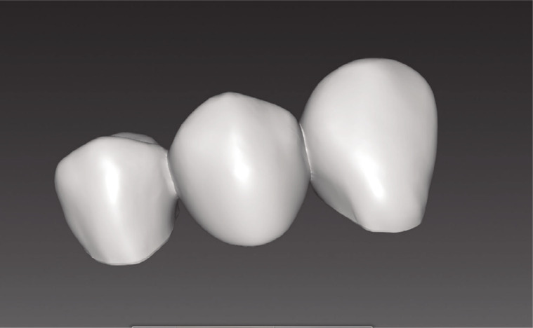
Materials and methods: For the fabrication of 3-unit monolithic zirconia restorations using impressions taken from the maxillary master cast, plaster cast was created and scanned in laboratory scanners (InEos X5 and D900L). The main cast was also scanned with different intraoral scanners (Omnicam [OMNI], Primescan [PS], Trios 3 [T3], Trios 4 [T4]) (n = 12 per group). Zirconia fixed partial dentures were virtually designed, produced from presintered block, and subsequently sintered. Marginal and internal discrepancy values (in µm) were measured by using silicone replica method under stereomicroscope. Data were statistically analyzed by using 1-way ANOVA and Kruskal Wallis tests (P<.05).
Results: In terms of marginal adaptation, the measurements on the canine tooth indicated better performance with intraoral scanners than those in laboratory scanners, but there was no difference among intraoral scanners (P<.05). In the premolar tooth, PS had the lowest marginal (86.9 ± 19.2 µm) and axial (92.4 ± 14.8 µm), and T4 had the lowest axio-occlusal (89.4 ± 15.6 µm) and occlusal (89.1 ± 13.9 µm) discrepancy value. In both canine and premolar teeth, the D900L was found to be the most marginally and internally inconsistent scanner.
Conclusion: Within the limits of the study, marginal and internal discrepancy values were generally lower in intraoral scanners than in laboratory scanners. Marginal discrepancy values of scanners were clinically acceptable (< 120 µm), except D900L.
期刊介绍:
This journal aims to convey scientific and clinical progress in the field of prosthodontics and its related areas to many dental communities concerned with esthetic and functional restorations, occlusion, implants, prostheses, and biomaterials related to prosthodontics.
This journal publishes
• Original research data of high scientific merit in the field of diagnosis, function, esthetics and stomatognathic physiology related to prosthodontic rehabilitation, physiology and mechanics of occlusion, mechanical and biologic aspects of prosthodontic materials including dental implants.
• Review articles by experts on controversies and new developments in prosthodontics.
• Case reports if they provide or document new fundamental knowledge.


 分享
分享
 求助内容:
求助内容: 应助结果提醒方式:
 扫码关注我们
