N S Martynenko, N Y Anisimova, M V Kiselevskiy, D R Temralieva, G I Raab, E A Kornyushenkov, M V Rodionov, S V Dobatkin, Y Z Estrin
{"title":"可吸收镁合金的体外生物降解研究。","authors":"N S Martynenko, N Y Anisimova, M V Kiselevskiy, D R Temralieva, G I Raab, E A Kornyushenkov, M V Rodionov, S V Dobatkin, Y Z Estrin","doi":"10.17691/stm2020.12.6.06","DOIUrl":null,"url":null,"abstract":"<p><p><b>The aim of the investigation</b> was to study the biodegradation characteristics and rate of magnesium alloys <i>in vitro</i>.</p><p><strong>Materials and methods: </strong>We studied the biodegradation of magnesium alloys Mg-Zn-Ca and WE43 (Mg-Y-Nd-Zr) in homogenized (initial) condition and after strengthening by mechanical processing using equal channel angular pressing (ECAP). The samples were incubated in a model system based on reference fetal calf serum (FCS) in the static and dynamic modes. The morphology of alloy surfaces was analyzed using light microscopy and computed tomography. Biodegradation was assessed by calculating weight loss within a certain incubation period. Cell adhesion and colonization stimulation were quantified in terms of a cell index (CI) using an analyzer xCELLigence RTCA Systems (ACEA Biosciences, Inc., USA) during the incubation of HEK 293 cells on WE43 specimens.</p><p><strong>Results: </strong>Strengthening of magnesium alloys Mg-Zn-Ca and WE43 using ECAP and, consequently, the changed structure resulted in the biodegradation acceleration as high as eightfold. Among the specimens incubated in FCS in different modes, those incubated in liquid flow exhibited the biodegradation rate twice as high as that of the specimens tested under static conditions. The biodegradation process was accompanied by local corrosion, although the degradation was primarily concentrated along the specimen margins stimulating cell adhesion and colonization. Such nature of degradation, as a rule, does not lead to anisotropy of the strength characteristics, that is important for medical materials. Superficial degradation of the alloys with no X-ray density changes in the bulk of the specimens was confirmed by computed tomography.</p><p><strong>Conclusion: </strong>The study of the biodegradation rate and further characteristics of magnesium alloys Mg-Zn-Ca and WE43 showed that the materials in both structural conditions are suitable for implants and can be used in bone implants and surgical fasteners.</p>","PeriodicalId":51886,"journal":{"name":"Sovremennye Tehnologii v Medicine","volume":"12 6","pages":"47-52"},"PeriodicalIF":0.9000,"publicationDate":"2021-01-01","publicationTypes":"Journal Article","fieldsOfStudy":null,"isOpenAccess":false,"openAccessPdf":"https://www.ncbi.nlm.nih.gov/pmc/articles/PMC8596234/pdf/","citationCount":"0","resultStr":"{\"title\":\"<i>In Vitro</i> Biodegradation of Resorbable Magnesium Alloys Promising for Implant Development.\",\"authors\":\"N S Martynenko, N Y Anisimova, M V Kiselevskiy, D R Temralieva, G I Raab, E A Kornyushenkov, M V Rodionov, S V Dobatkin, Y Z Estrin\",\"doi\":\"10.17691/stm2020.12.6.06\",\"DOIUrl\":null,\"url\":null,\"abstract\":\"<p><p><b>The aim of the investigation</b> was to study the biodegradation characteristics and rate of magnesium alloys <i>in vitro</i>.</p><p><strong>Materials and methods: </strong>We studied the biodegradation of magnesium alloys Mg-Zn-Ca and WE43 (Mg-Y-Nd-Zr) in homogenized (initial) condition and after strengthening by mechanical processing using equal channel angular pressing (ECAP). The samples were incubated in a model system based on reference fetal calf serum (FCS) in the static and dynamic modes. The morphology of alloy surfaces was analyzed using light microscopy and computed tomography. Biodegradation was assessed by calculating weight loss within a certain incubation period. Cell adhesion and colonization stimulation were quantified in terms of a cell index (CI) using an analyzer xCELLigence RTCA Systems (ACEA Biosciences, Inc., USA) during the incubation of HEK 293 cells on WE43 specimens.</p><p><strong>Results: </strong>Strengthening of magnesium alloys Mg-Zn-Ca and WE43 using ECAP and, consequently, the changed structure resulted in the biodegradation acceleration as high as eightfold. Among the specimens incubated in FCS in different modes, those incubated in liquid flow exhibited the biodegradation rate twice as high as that of the specimens tested under static conditions. The biodegradation process was accompanied by local corrosion, although the degradation was primarily concentrated along the specimen margins stimulating cell adhesion and colonization. Such nature of degradation, as a rule, does not lead to anisotropy of the strength characteristics, that is important for medical materials. Superficial degradation of the alloys with no X-ray density changes in the bulk of the specimens was confirmed by computed tomography.</p><p><strong>Conclusion: </strong>The study of the biodegradation rate and further characteristics of magnesium alloys Mg-Zn-Ca and WE43 showed that the materials in both structural conditions are suitable for implants and can be used in bone implants and surgical fasteners.</p>\",\"PeriodicalId\":51886,\"journal\":{\"name\":\"Sovremennye Tehnologii v Medicine\",\"volume\":\"12 6\",\"pages\":\"47-52\"},\"PeriodicalIF\":0.9000,\"publicationDate\":\"2021-01-01\",\"publicationTypes\":\"Journal Article\",\"fieldsOfStudy\":null,\"isOpenAccess\":false,\"openAccessPdf\":\"https://www.ncbi.nlm.nih.gov/pmc/articles/PMC8596234/pdf/\",\"citationCount\":\"0\",\"resultStr\":null,\"platform\":\"Semanticscholar\",\"paperid\":null,\"PeriodicalName\":\"Sovremennye Tehnologii v Medicine\",\"FirstCategoryId\":\"1085\",\"ListUrlMain\":\"https://doi.org/10.17691/stm2020.12.6.06\",\"RegionNum\":0,\"RegionCategory\":null,\"ArticlePicture\":[],\"TitleCN\":null,\"AbstractTextCN\":null,\"PMCID\":null,\"EPubDate\":\"2020/12/28 0:00:00\",\"PubModel\":\"Epub\",\"JCR\":\"Q4\",\"JCRName\":\"MEDICINE, RESEARCH & EXPERIMENTAL\",\"Score\":null,\"Total\":0}","platform":"Semanticscholar","paperid":null,"PeriodicalName":"Sovremennye Tehnologii v Medicine","FirstCategoryId":"1085","ListUrlMain":"https://doi.org/10.17691/stm2020.12.6.06","RegionNum":0,"RegionCategory":null,"ArticlePicture":[],"TitleCN":null,"AbstractTextCN":null,"PMCID":null,"EPubDate":"2020/12/28 0:00:00","PubModel":"Epub","JCR":"Q4","JCRName":"MEDICINE, RESEARCH & EXPERIMENTAL","Score":null,"Total":0}
引用次数: 0
摘要
研究镁合金在体外的生物降解特性及降解速率。材料与方法:研究了Mg-Zn-Ca和WE43镁合金(Mg-Y-Nd-Zr)在均质(初始)和等通道角压(ECAP)强化后的生物降解。样品在以参考胎牛血清(FCS)为基础的模型系统中进行静态和动态培养。采用光学显微镜和计算机断层扫描对合金表面形貌进行了分析。通过计算一定潜伏期内的失重来评估生物降解。在HEK 293细胞在WE43标本上孵育期间,使用xCELLigence RTCA Systems (ACEA Biosciences, Inc., USA)分析仪,以细胞指数(CI)定量细胞粘附和定植刺激。结果:ECAP对Mg-Zn-Ca镁合金和WE43镁合金进行强化,使其结构发生改变,生物降解加速高达8倍。在FCS中不同模式培养的样品中,液体流动培养的样品的生物降解率是静态条件下的两倍。生物降解过程伴随着局部腐蚀,尽管降解主要集中在刺激细胞粘附和定植的标本边缘。通常,这种降解性质不会导致强度特性的各向异性,这对医用材料很重要。通过计算机断层扫描证实了合金的表面降解,但大部分试样的x射线密度没有变化。结论:对Mg-Zn-Ca镁合金和WE43镁合金的生物降解率及进一步特性的研究表明,两种结构条件下的材料均适用于种植体,可用于骨种植体和外科固定物。
In Vitro Biodegradation of Resorbable Magnesium Alloys Promising for Implant Development.
The aim of the investigation was to study the biodegradation characteristics and rate of magnesium alloys in vitro.
Materials and methods: We studied the biodegradation of magnesium alloys Mg-Zn-Ca and WE43 (Mg-Y-Nd-Zr) in homogenized (initial) condition and after strengthening by mechanical processing using equal channel angular pressing (ECAP). The samples were incubated in a model system based on reference fetal calf serum (FCS) in the static and dynamic modes. The morphology of alloy surfaces was analyzed using light microscopy and computed tomography. Biodegradation was assessed by calculating weight loss within a certain incubation period. Cell adhesion and colonization stimulation were quantified in terms of a cell index (CI) using an analyzer xCELLigence RTCA Systems (ACEA Biosciences, Inc., USA) during the incubation of HEK 293 cells on WE43 specimens.
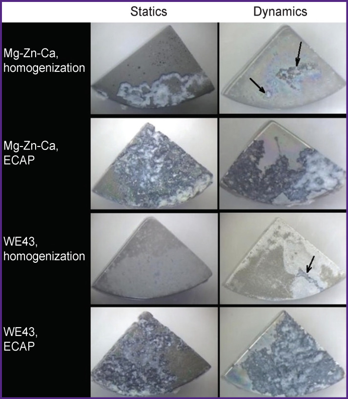
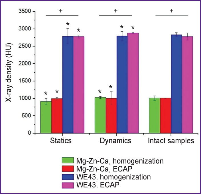
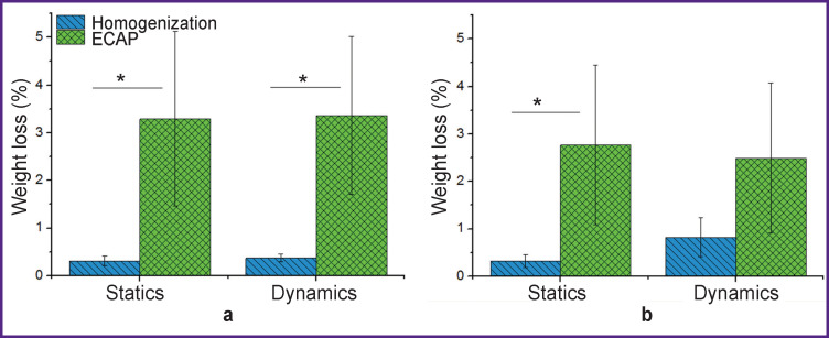
Results: Strengthening of magnesium alloys Mg-Zn-Ca and WE43 using ECAP and, consequently, the changed structure resulted in the biodegradation acceleration as high as eightfold. Among the specimens incubated in FCS in different modes, those incubated in liquid flow exhibited the biodegradation rate twice as high as that of the specimens tested under static conditions. The biodegradation process was accompanied by local corrosion, although the degradation was primarily concentrated along the specimen margins stimulating cell adhesion and colonization. Such nature of degradation, as a rule, does not lead to anisotropy of the strength characteristics, that is important for medical materials. Superficial degradation of the alloys with no X-ray density changes in the bulk of the specimens was confirmed by computed tomography.
Conclusion: The study of the biodegradation rate and further characteristics of magnesium alloys Mg-Zn-Ca and WE43 showed that the materials in both structural conditions are suitable for implants and can be used in bone implants and surgical fasteners.


 分享
分享
 求助内容:
求助内容: 应助结果提醒方式:
 扫码关注我们
