Adam J. Blanch , Juan Nunez-Iglesias , Arman Namvar , Sebastien Menant , Oliver Looker , Vijay Rajagopal , Wai-Hong Tham , Leann Tilley , Matthew W.A. Dixon
{"title":"多模态成像显示网状红细胞成熟过程中的膜骨架重组以及成熟红细胞的窝区和边缘区差异","authors":"Adam J. Blanch , Juan Nunez-Iglesias , Arman Namvar , Sebastien Menant , Oliver Looker , Vijay Rajagopal , Wai-Hong Tham , Leann Tilley , Matthew W.A. Dixon","doi":"10.1016/j.yjsbx.2021.100056","DOIUrl":null,"url":null,"abstract":"<div><p>The red blood cell (RBC) is remarkable in its ability to deform as it passages through the vasculature. Its deformability derives from a spectrin-actin protein network that supports the cell membrane and provides strength and flexibility, however questions remain regarding the assembly and maintenance of the skeletal network. Using scanning electron microscopy (SEM) and atomic force microscopy (AFM) we have examined the nanoscale architecture of the cytoplasmic side of membrane discs prepared from reticulocytes and mature RBCs. Immunofluorescence microscopy was used to probe the distribution of spectrin and other membrane skeleton proteins. We found that the cell surface area decreases by up to 30% and the spectrin-actin network increases in density by approximately 20% as the reticulocyte matures. By contrast, the inter-junctional distance and junctional density increase only by 3–4% and 5–9%, respectively. This suggests that the maturation-associated reduction in surface area is accompanied by an increase in spectrin self-association to form higher order oligomers. We also examined the mature RBC membrane in the edge (rim) and face (dimple) regions of mature RBCs and found the rim contains about 1.5% more junctional complexes compared to the dimple region. A 2% increase in band 4.1 density in the rim supports these structural measurements.</p></div>","PeriodicalId":17238,"journal":{"name":"Journal of Structural Biology: X","volume":"6 ","pages":"Article 100056"},"PeriodicalIF":5.1000,"publicationDate":"2022-01-01","publicationTypes":"Journal Article","fieldsOfStudy":null,"isOpenAccess":false,"openAccessPdf":"https://ftp.ncbi.nlm.nih.gov/pub/pmc/oa_pdf/80/3d/main.PMC8688873.pdf","citationCount":"2","resultStr":"{\"title\":\"Multimodal imaging reveals membrane skeleton reorganisation during reticulocyte maturation and differences in dimple and rim regions of mature erythrocytes\",\"authors\":\"Adam J. Blanch , Juan Nunez-Iglesias , Arman Namvar , Sebastien Menant , Oliver Looker , Vijay Rajagopal , Wai-Hong Tham , Leann Tilley , Matthew W.A. Dixon\",\"doi\":\"10.1016/j.yjsbx.2021.100056\",\"DOIUrl\":null,\"url\":null,\"abstract\":\"<div><p>The red blood cell (RBC) is remarkable in its ability to deform as it passages through the vasculature. Its deformability derives from a spectrin-actin protein network that supports the cell membrane and provides strength and flexibility, however questions remain regarding the assembly and maintenance of the skeletal network. Using scanning electron microscopy (SEM) and atomic force microscopy (AFM) we have examined the nanoscale architecture of the cytoplasmic side of membrane discs prepared from reticulocytes and mature RBCs. Immunofluorescence microscopy was used to probe the distribution of spectrin and other membrane skeleton proteins. We found that the cell surface area decreases by up to 30% and the spectrin-actin network increases in density by approximately 20% as the reticulocyte matures. By contrast, the inter-junctional distance and junctional density increase only by 3–4% and 5–9%, respectively. This suggests that the maturation-associated reduction in surface area is accompanied by an increase in spectrin self-association to form higher order oligomers. We also examined the mature RBC membrane in the edge (rim) and face (dimple) regions of mature RBCs and found the rim contains about 1.5% more junctional complexes compared to the dimple region. A 2% increase in band 4.1 density in the rim supports these structural measurements.</p></div>\",\"PeriodicalId\":17238,\"journal\":{\"name\":\"Journal of Structural Biology: X\",\"volume\":\"6 \",\"pages\":\"Article 100056\"},\"PeriodicalIF\":5.1000,\"publicationDate\":\"2022-01-01\",\"publicationTypes\":\"Journal Article\",\"fieldsOfStudy\":null,\"isOpenAccess\":false,\"openAccessPdf\":\"https://ftp.ncbi.nlm.nih.gov/pub/pmc/oa_pdf/80/3d/main.PMC8688873.pdf\",\"citationCount\":\"2\",\"resultStr\":null,\"platform\":\"Semanticscholar\",\"paperid\":null,\"PeriodicalName\":\"Journal of Structural Biology: X\",\"FirstCategoryId\":\"1085\",\"ListUrlMain\":\"https://www.sciencedirect.com/science/article/pii/S2590152421000131\",\"RegionNum\":0,\"RegionCategory\":null,\"ArticlePicture\":[],\"TitleCN\":null,\"AbstractTextCN\":null,\"PMCID\":null,\"EPubDate\":\"\",\"PubModel\":\"\",\"JCR\":\"Q2\",\"JCRName\":\"BIOCHEMISTRY & MOLECULAR BIOLOGY\",\"Score\":null,\"Total\":0}","platform":"Semanticscholar","paperid":null,"PeriodicalName":"Journal of Structural Biology: X","FirstCategoryId":"1085","ListUrlMain":"https://www.sciencedirect.com/science/article/pii/S2590152421000131","RegionNum":0,"RegionCategory":null,"ArticlePicture":[],"TitleCN":null,"AbstractTextCN":null,"PMCID":null,"EPubDate":"","PubModel":"","JCR":"Q2","JCRName":"BIOCHEMISTRY & MOLECULAR BIOLOGY","Score":null,"Total":0}
Multimodal imaging reveals membrane skeleton reorganisation during reticulocyte maturation and differences in dimple and rim regions of mature erythrocytes
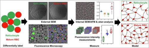
The red blood cell (RBC) is remarkable in its ability to deform as it passages through the vasculature. Its deformability derives from a spectrin-actin protein network that supports the cell membrane and provides strength and flexibility, however questions remain regarding the assembly and maintenance of the skeletal network. Using scanning electron microscopy (SEM) and atomic force microscopy (AFM) we have examined the nanoscale architecture of the cytoplasmic side of membrane discs prepared from reticulocytes and mature RBCs. Immunofluorescence microscopy was used to probe the distribution of spectrin and other membrane skeleton proteins. We found that the cell surface area decreases by up to 30% and the spectrin-actin network increases in density by approximately 20% as the reticulocyte matures. By contrast, the inter-junctional distance and junctional density increase only by 3–4% and 5–9%, respectively. This suggests that the maturation-associated reduction in surface area is accompanied by an increase in spectrin self-association to form higher order oligomers. We also examined the mature RBC membrane in the edge (rim) and face (dimple) regions of mature RBCs and found the rim contains about 1.5% more junctional complexes compared to the dimple region. A 2% increase in band 4.1 density in the rim supports these structural measurements.


 分享
分享
 求助内容:
求助内容: 应助结果提醒方式:
 扫码关注我们
