{"title":"微距摄影与光片照明使整个扩展脑成像单细胞分辨率。","authors":"Chia-Ming Lee, Xuejiao Tian, Chieh Tsao, Peilin Chen, Tzyy-Nan Huang, Yi-Ping Hsueh, Bi-Chang Chen","doi":"10.15190/d.2021.12","DOIUrl":null,"url":null,"abstract":"<p><p>Macro photography allows direct visualization of the enlarged whole mouse brain by a combination of lightsheet illumination and expansion microscopy with single-cell resolution. Taking advantage of the long working distance of a camera lens, we imaged a 3.7 cm thick, transparent, fluorescently-labeled expanded brain. In order to improve 3D sectioning capability, we used lightsheet excitation confined as the depth of field of the camera lens. Using 4x sample expansion and 5x optical magnification, macro photography enables imaging of expanded whole mouse brain with an effective resolution of 300 nm, which provides the subcellular structural information at the organ level.</p>","PeriodicalId":72829,"journal":{"name":"Discoveries (Craiova, Romania)","volume":null,"pages":null},"PeriodicalIF":0.0000,"publicationDate":"2021-08-04","publicationTypes":"Journal Article","fieldsOfStudy":null,"isOpenAccess":false,"openAccessPdf":"https://www.ncbi.nlm.nih.gov/pmc/articles/PMC8626140/pdf/","citationCount":"1","resultStr":"{\"title\":\"Macro Photography with Lightsheet Illumination Enables Whole Expanded Brain Imaging with Single-cell Resolution.\",\"authors\":\"Chia-Ming Lee, Xuejiao Tian, Chieh Tsao, Peilin Chen, Tzyy-Nan Huang, Yi-Ping Hsueh, Bi-Chang Chen\",\"doi\":\"10.15190/d.2021.12\",\"DOIUrl\":null,\"url\":null,\"abstract\":\"<p><p>Macro photography allows direct visualization of the enlarged whole mouse brain by a combination of lightsheet illumination and expansion microscopy with single-cell resolution. Taking advantage of the long working distance of a camera lens, we imaged a 3.7 cm thick, transparent, fluorescently-labeled expanded brain. In order to improve 3D sectioning capability, we used lightsheet excitation confined as the depth of field of the camera lens. Using 4x sample expansion and 5x optical magnification, macro photography enables imaging of expanded whole mouse brain with an effective resolution of 300 nm, which provides the subcellular structural information at the organ level.</p>\",\"PeriodicalId\":72829,\"journal\":{\"name\":\"Discoveries (Craiova, Romania)\",\"volume\":null,\"pages\":null},\"PeriodicalIF\":0.0000,\"publicationDate\":\"2021-08-04\",\"publicationTypes\":\"Journal Article\",\"fieldsOfStudy\":null,\"isOpenAccess\":false,\"openAccessPdf\":\"https://www.ncbi.nlm.nih.gov/pmc/articles/PMC8626140/pdf/\",\"citationCount\":\"1\",\"resultStr\":null,\"platform\":\"Semanticscholar\",\"paperid\":null,\"PeriodicalName\":\"Discoveries (Craiova, Romania)\",\"FirstCategoryId\":\"1085\",\"ListUrlMain\":\"https://doi.org/10.15190/d.2021.12\",\"RegionNum\":0,\"RegionCategory\":null,\"ArticlePicture\":[],\"TitleCN\":null,\"AbstractTextCN\":null,\"PMCID\":null,\"EPubDate\":\"2021/7/1 0:00:00\",\"PubModel\":\"eCollection\",\"JCR\":\"\",\"JCRName\":\"\",\"Score\":null,\"Total\":0}","platform":"Semanticscholar","paperid":null,"PeriodicalName":"Discoveries (Craiova, Romania)","FirstCategoryId":"1085","ListUrlMain":"https://doi.org/10.15190/d.2021.12","RegionNum":0,"RegionCategory":null,"ArticlePicture":[],"TitleCN":null,"AbstractTextCN":null,"PMCID":null,"EPubDate":"2021/7/1 0:00:00","PubModel":"eCollection","JCR":"","JCRName":"","Score":null,"Total":0}
Macro Photography with Lightsheet Illumination Enables Whole Expanded Brain Imaging with Single-cell Resolution.
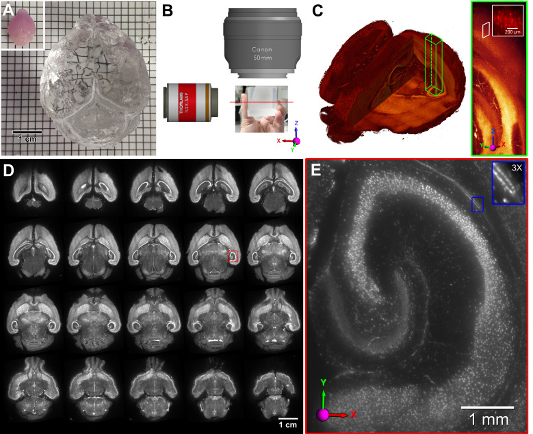
Macro photography allows direct visualization of the enlarged whole mouse brain by a combination of lightsheet illumination and expansion microscopy with single-cell resolution. Taking advantage of the long working distance of a camera lens, we imaged a 3.7 cm thick, transparent, fluorescently-labeled expanded brain. In order to improve 3D sectioning capability, we used lightsheet excitation confined as the depth of field of the camera lens. Using 4x sample expansion and 5x optical magnification, macro photography enables imaging of expanded whole mouse brain with an effective resolution of 300 nm, which provides the subcellular structural information at the organ level.


 分享
分享

 求助内容:
求助内容: 应助结果提醒方式:
 扫码关注我们
