{"title":"双肺弥漫性氟脱氧葡萄糖摄取:低剂量CT表现支持的过敏性肺炎。","authors":"Shun Goto, Yohji Matsusaka, Tomohiko Yamane, Yuki Hoshino, Ichiei Kuji","doi":"10.22038/AOJNMB.2021.56000.1393","DOIUrl":null,"url":null,"abstract":"<p><p>Hypersensitivity pneumonitis (HP) is an interstitial lung disease resulting from an immune-mediated response in susceptible and sensitized individuals to various inhaled antigens in the environment. Imaging diagnosis is usually based on high-resolution CT findings. Here, we present a 49-year-old man with a history of diffuse large B-cell lymphoma presented with fever and occasional cough. <sup>18</sup>F-fluorodeoxyglucose (FDG) positron emission tomography/computed tomography (PET/CT) revealed diffuse FDG uptake in the bilateral lungs. Expiratory low-dose CT simultaneously performed in PET scanning revealed centrilobular nodules and air trapping in ground glass opacities (GGO). Our imaging diagnosis was acute hypersensitivity pneumonitis (HP). Based on the results of his clinical course, blood laboratory tests, and bronchoscopy, he was diagnosed with acute HP. Diffuse pulmonary FDG uptake can be seen in the patients with acute HP. In addition, expiratory low-dose CT findings of centrilobular nodules and air trapping in GGO may be helpful for accurate diagnosis of acute HP.</p>","PeriodicalId":8503,"journal":{"name":"Asia Oceania Journal of Nuclear Medicine and Biology","volume":"10 1","pages":"43-46"},"PeriodicalIF":0.0000,"publicationDate":"2022-01-01","publicationTypes":"Journal Article","fieldsOfStudy":null,"isOpenAccess":false,"openAccessPdf":"https://www.ncbi.nlm.nih.gov/pmc/articles/PMC8742853/pdf/","citationCount":"0","resultStr":"{\"title\":\"Diffuse FDG uptake in the bilateral lungs: hypersensitivity pneumonitis supported by low-dose CT findings.\",\"authors\":\"Shun Goto, Yohji Matsusaka, Tomohiko Yamane, Yuki Hoshino, Ichiei Kuji\",\"doi\":\"10.22038/AOJNMB.2021.56000.1393\",\"DOIUrl\":null,\"url\":null,\"abstract\":\"<p><p>Hypersensitivity pneumonitis (HP) is an interstitial lung disease resulting from an immune-mediated response in susceptible and sensitized individuals to various inhaled antigens in the environment. Imaging diagnosis is usually based on high-resolution CT findings. Here, we present a 49-year-old man with a history of diffuse large B-cell lymphoma presented with fever and occasional cough. <sup>18</sup>F-fluorodeoxyglucose (FDG) positron emission tomography/computed tomography (PET/CT) revealed diffuse FDG uptake in the bilateral lungs. Expiratory low-dose CT simultaneously performed in PET scanning revealed centrilobular nodules and air trapping in ground glass opacities (GGO). Our imaging diagnosis was acute hypersensitivity pneumonitis (HP). Based on the results of his clinical course, blood laboratory tests, and bronchoscopy, he was diagnosed with acute HP. Diffuse pulmonary FDG uptake can be seen in the patients with acute HP. In addition, expiratory low-dose CT findings of centrilobular nodules and air trapping in GGO may be helpful for accurate diagnosis of acute HP.</p>\",\"PeriodicalId\":8503,\"journal\":{\"name\":\"Asia Oceania Journal of Nuclear Medicine and Biology\",\"volume\":\"10 1\",\"pages\":\"43-46\"},\"PeriodicalIF\":0.0000,\"publicationDate\":\"2022-01-01\",\"publicationTypes\":\"Journal Article\",\"fieldsOfStudy\":null,\"isOpenAccess\":false,\"openAccessPdf\":\"https://www.ncbi.nlm.nih.gov/pmc/articles/PMC8742853/pdf/\",\"citationCount\":\"0\",\"resultStr\":null,\"platform\":\"Semanticscholar\",\"paperid\":null,\"PeriodicalName\":\"Asia Oceania Journal of Nuclear Medicine and Biology\",\"FirstCategoryId\":\"1085\",\"ListUrlMain\":\"https://doi.org/10.22038/AOJNMB.2021.56000.1393\",\"RegionNum\":0,\"RegionCategory\":null,\"ArticlePicture\":[],\"TitleCN\":null,\"AbstractTextCN\":null,\"PMCID\":null,\"EPubDate\":\"\",\"PubModel\":\"\",\"JCR\":\"Q3\",\"JCRName\":\"Medicine\",\"Score\":null,\"Total\":0}","platform":"Semanticscholar","paperid":null,"PeriodicalName":"Asia Oceania Journal of Nuclear Medicine and Biology","FirstCategoryId":"1085","ListUrlMain":"https://doi.org/10.22038/AOJNMB.2021.56000.1393","RegionNum":0,"RegionCategory":null,"ArticlePicture":[],"TitleCN":null,"AbstractTextCN":null,"PMCID":null,"EPubDate":"","PubModel":"","JCR":"Q3","JCRName":"Medicine","Score":null,"Total":0}
Diffuse FDG uptake in the bilateral lungs: hypersensitivity pneumonitis supported by low-dose CT findings.
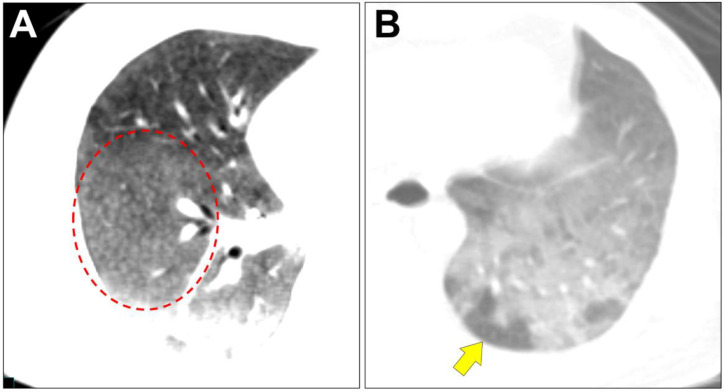
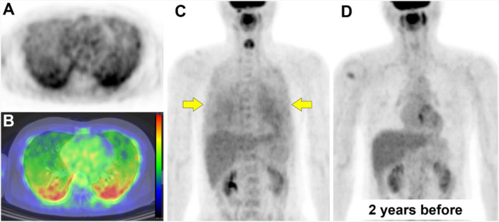
Hypersensitivity pneumonitis (HP) is an interstitial lung disease resulting from an immune-mediated response in susceptible and sensitized individuals to various inhaled antigens in the environment. Imaging diagnosis is usually based on high-resolution CT findings. Here, we present a 49-year-old man with a history of diffuse large B-cell lymphoma presented with fever and occasional cough. 18F-fluorodeoxyglucose (FDG) positron emission tomography/computed tomography (PET/CT) revealed diffuse FDG uptake in the bilateral lungs. Expiratory low-dose CT simultaneously performed in PET scanning revealed centrilobular nodules and air trapping in ground glass opacities (GGO). Our imaging diagnosis was acute hypersensitivity pneumonitis (HP). Based on the results of his clinical course, blood laboratory tests, and bronchoscopy, he was diagnosed with acute HP. Diffuse pulmonary FDG uptake can be seen in the patients with acute HP. In addition, expiratory low-dose CT findings of centrilobular nodules and air trapping in GGO may be helpful for accurate diagnosis of acute HP.


 分享
分享
 求助内容:
求助内容: 应助结果提醒方式:
 扫码关注我们
