{"title":"急性前循环缺血性脑卒中的成像:当前和未来。","authors":"Hyun Jeong Kim, Hong Gee Roh","doi":"10.5469/neuroint.2021.00465","DOIUrl":null,"url":null,"abstract":"<p><p>Clinical trials on acute ischemic stroke have demonstrated the clinical effectiveness of revascularization treatments within an appropriate time window after stroke onset: intravenous thrombolysis (NINDS and ECASS-III) through the administration of tissue plasminogen activator within a 4.5-hour time window, endovascular thrombectomy (ESCAPE, REVASCAT, SWIFT-PRIME, MR CLEAN, EXTEND-IA) within a 6-hour time window, and extending the treatment time window up to 24 hours for endovascular thrombectomy (DAWN and DEFUSE 3). However, a substantial number of patients in these trials were ineligible for revascularization treatment, and treatments of some patients were considerably futile or sometimes dangerous in the clinical trials. Guidelines for the early management of patients with acute ischemic stroke have evolved to accept revascularization treatment as standard and include eligibility criteria for the treatment. Imaging has been crucial in selecting eligible patients for revascularization treatment in guidelines and clinical trials. Stroke specialists should know imaging criteria for revascularization treatment. Stroke imaging studies have demonstrated imaging roles in acute ischemic stroke management as follows: 1) exclusion of hemorrhage and stroke mimic disease, 2) assessment of salvageable brain, 3) localization of the site of vascular occlusion and thrombus, 4) estimation of collateral circulation, and 5) prediction of acute ischemic stroke expecting hemorrhagic transformation. Here, we review imaging methods and criteria to select eligible patients for revascularization treatment in acute anterior circulation stroke, focus on 2019 guidelines from the American Heart Association/American Stroke Association, and discuss the future direction of imaging-based patient selection to improve treatment effects.</p>","PeriodicalId":19140,"journal":{"name":"Neurointervention","volume":"17 1","pages":"2-17"},"PeriodicalIF":1.2000,"publicationDate":"2022-03-01","publicationTypes":"Journal Article","fieldsOfStudy":null,"isOpenAccess":false,"openAccessPdf":"https://ftp.ncbi.nlm.nih.gov/pub/pmc/oa_pdf/27/3b/neuroint-2021-00465.PMC8891584.pdf","citationCount":"0","resultStr":"{\"title\":\"Imaging in Acute Anterior Circulation Ischemic Stroke: Current and Future.\",\"authors\":\"Hyun Jeong Kim, Hong Gee Roh\",\"doi\":\"10.5469/neuroint.2021.00465\",\"DOIUrl\":null,\"url\":null,\"abstract\":\"<p><p>Clinical trials on acute ischemic stroke have demonstrated the clinical effectiveness of revascularization treatments within an appropriate time window after stroke onset: intravenous thrombolysis (NINDS and ECASS-III) through the administration of tissue plasminogen activator within a 4.5-hour time window, endovascular thrombectomy (ESCAPE, REVASCAT, SWIFT-PRIME, MR CLEAN, EXTEND-IA) within a 6-hour time window, and extending the treatment time window up to 24 hours for endovascular thrombectomy (DAWN and DEFUSE 3). However, a substantial number of patients in these trials were ineligible for revascularization treatment, and treatments of some patients were considerably futile or sometimes dangerous in the clinical trials. Guidelines for the early management of patients with acute ischemic stroke have evolved to accept revascularization treatment as standard and include eligibility criteria for the treatment. Imaging has been crucial in selecting eligible patients for revascularization treatment in guidelines and clinical trials. Stroke specialists should know imaging criteria for revascularization treatment. Stroke imaging studies have demonstrated imaging roles in acute ischemic stroke management as follows: 1) exclusion of hemorrhage and stroke mimic disease, 2) assessment of salvageable brain, 3) localization of the site of vascular occlusion and thrombus, 4) estimation of collateral circulation, and 5) prediction of acute ischemic stroke expecting hemorrhagic transformation. Here, we review imaging methods and criteria to select eligible patients for revascularization treatment in acute anterior circulation stroke, focus on 2019 guidelines from the American Heart Association/American Stroke Association, and discuss the future direction of imaging-based patient selection to improve treatment effects.</p>\",\"PeriodicalId\":19140,\"journal\":{\"name\":\"Neurointervention\",\"volume\":\"17 1\",\"pages\":\"2-17\"},\"PeriodicalIF\":1.2000,\"publicationDate\":\"2022-03-01\",\"publicationTypes\":\"Journal Article\",\"fieldsOfStudy\":null,\"isOpenAccess\":false,\"openAccessPdf\":\"https://ftp.ncbi.nlm.nih.gov/pub/pmc/oa_pdf/27/3b/neuroint-2021-00465.PMC8891584.pdf\",\"citationCount\":\"0\",\"resultStr\":null,\"platform\":\"Semanticscholar\",\"paperid\":null,\"PeriodicalName\":\"Neurointervention\",\"FirstCategoryId\":\"1085\",\"ListUrlMain\":\"https://doi.org/10.5469/neuroint.2021.00465\",\"RegionNum\":0,\"RegionCategory\":null,\"ArticlePicture\":[],\"TitleCN\":null,\"AbstractTextCN\":null,\"PMCID\":null,\"EPubDate\":\"2022/2/4 0:00:00\",\"PubModel\":\"Epub\",\"JCR\":\"Q4\",\"JCRName\":\"CLINICAL NEUROLOGY\",\"Score\":null,\"Total\":0}","platform":"Semanticscholar","paperid":null,"PeriodicalName":"Neurointervention","FirstCategoryId":"1085","ListUrlMain":"https://doi.org/10.5469/neuroint.2021.00465","RegionNum":0,"RegionCategory":null,"ArticlePicture":[],"TitleCN":null,"AbstractTextCN":null,"PMCID":null,"EPubDate":"2022/2/4 0:00:00","PubModel":"Epub","JCR":"Q4","JCRName":"CLINICAL NEUROLOGY","Score":null,"Total":0}
Imaging in Acute Anterior Circulation Ischemic Stroke: Current and Future.
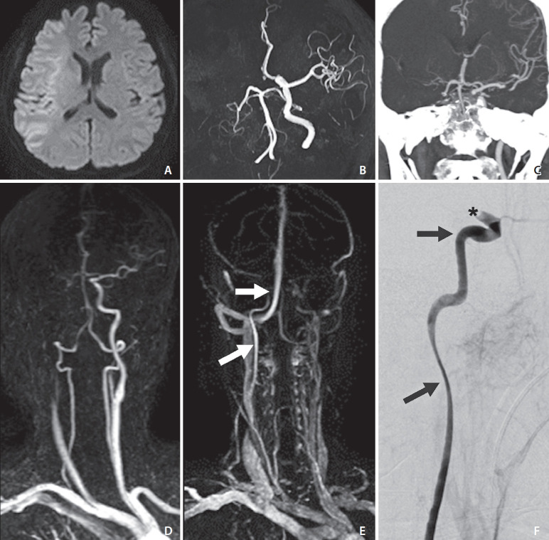
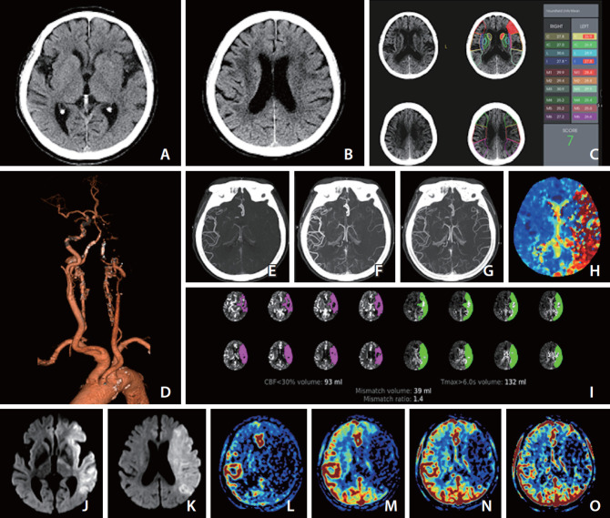
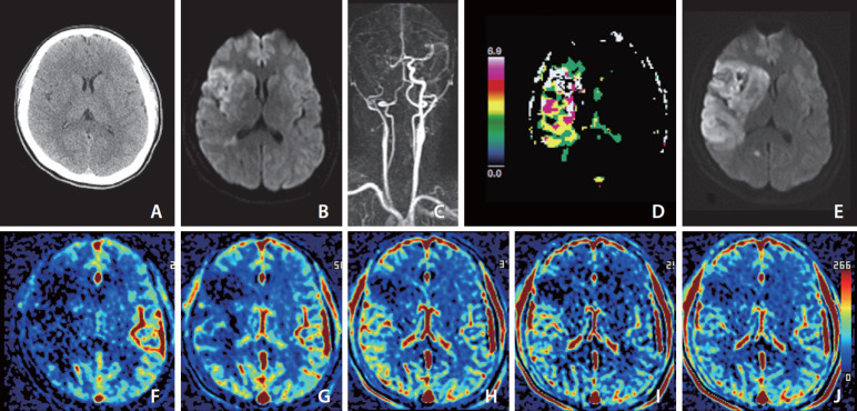
Clinical trials on acute ischemic stroke have demonstrated the clinical effectiveness of revascularization treatments within an appropriate time window after stroke onset: intravenous thrombolysis (NINDS and ECASS-III) through the administration of tissue plasminogen activator within a 4.5-hour time window, endovascular thrombectomy (ESCAPE, REVASCAT, SWIFT-PRIME, MR CLEAN, EXTEND-IA) within a 6-hour time window, and extending the treatment time window up to 24 hours for endovascular thrombectomy (DAWN and DEFUSE 3). However, a substantial number of patients in these trials were ineligible for revascularization treatment, and treatments of some patients were considerably futile or sometimes dangerous in the clinical trials. Guidelines for the early management of patients with acute ischemic stroke have evolved to accept revascularization treatment as standard and include eligibility criteria for the treatment. Imaging has been crucial in selecting eligible patients for revascularization treatment in guidelines and clinical trials. Stroke specialists should know imaging criteria for revascularization treatment. Stroke imaging studies have demonstrated imaging roles in acute ischemic stroke management as follows: 1) exclusion of hemorrhage and stroke mimic disease, 2) assessment of salvageable brain, 3) localization of the site of vascular occlusion and thrombus, 4) estimation of collateral circulation, and 5) prediction of acute ischemic stroke expecting hemorrhagic transformation. Here, we review imaging methods and criteria to select eligible patients for revascularization treatment in acute anterior circulation stroke, focus on 2019 guidelines from the American Heart Association/American Stroke Association, and discuss the future direction of imaging-based patient selection to improve treatment effects.


 分享
分享

 求助内容:
求助内容: 应助结果提醒方式:
 扫码关注我们
