Thomas Keating , Samuel Lethbridge , Jon C. Allnutt , Charlotte L. Hendon-Dunn , Stephen R. Thomas , Luke J. Alderwick , Stephen C. Taylor , Joanna Bacon
{"title":"结核分枝杆菌在生物膜生长过程中改变细胞壁碳水化合物,同时降低补体激活","authors":"Thomas Keating , Samuel Lethbridge , Jon C. Allnutt , Charlotte L. Hendon-Dunn , Stephen R. Thomas , Luke J. Alderwick , Stephen C. Taylor , Joanna Bacon","doi":"10.1016/j.tcsw.2021.100065","DOIUrl":null,"url":null,"abstract":"<div><p>The development of new vaccines for TB needs to be underpinned by an understanding of both the molecular and cellular mechanisms of host-pathogen interactions and how the immune response can be modulated to achieve protection from disease. Complement orchestrates many aspects of the innate and adaptive immune responses. However, little is known about the contribution of the complement pathways during TB disease, particularly with respect to mycobacterial phenotype. Extracellular communities (biofilms) of <em>M. tuberculosis</em> are found in the acellular rim of granulomas, during disease, and these are likely to be present in post-primary TB episodes, in necrotic lesions. Our study aimed to determine which mycobacterial cell wall components were altered during biofilm growth and how these cell wall alterations modified the complement response. We have shown that <em>M. tuberculosis</em> biofilms modified their cell wall carbohydrates and elicited reduced classical and lectin pathway activation. Consistent with this finding was the reduction of C3b/iC3b deposition on biofilm cell wall carbohydrate extracts. Here, we have highlighted the role of cell wall carbohydrate alterations during biofilm growth of <em>M. tuberculosis</em> and subsequent modulation of complement activation.</p></div>","PeriodicalId":36539,"journal":{"name":"Cell Surface","volume":"7 ","pages":"Article 100065"},"PeriodicalIF":6.2000,"publicationDate":"2021-12-01","publicationTypes":"Journal Article","fieldsOfStudy":null,"isOpenAccess":false,"openAccessPdf":"https://ftp.ncbi.nlm.nih.gov/pub/pmc/oa_pdf/3a/4a/main.PMC8577165.pdf","citationCount":"2","resultStr":"{\"title\":\"Mycobacterium tuberculosis modifies cell wall carbohydrates during biofilm growth with a concomitant reduction in complement activation\",\"authors\":\"Thomas Keating , Samuel Lethbridge , Jon C. Allnutt , Charlotte L. Hendon-Dunn , Stephen R. Thomas , Luke J. Alderwick , Stephen C. Taylor , Joanna Bacon\",\"doi\":\"10.1016/j.tcsw.2021.100065\",\"DOIUrl\":null,\"url\":null,\"abstract\":\"<div><p>The development of new vaccines for TB needs to be underpinned by an understanding of both the molecular and cellular mechanisms of host-pathogen interactions and how the immune response can be modulated to achieve protection from disease. Complement orchestrates many aspects of the innate and adaptive immune responses. However, little is known about the contribution of the complement pathways during TB disease, particularly with respect to mycobacterial phenotype. Extracellular communities (biofilms) of <em>M. tuberculosis</em> are found in the acellular rim of granulomas, during disease, and these are likely to be present in post-primary TB episodes, in necrotic lesions. Our study aimed to determine which mycobacterial cell wall components were altered during biofilm growth and how these cell wall alterations modified the complement response. We have shown that <em>M. tuberculosis</em> biofilms modified their cell wall carbohydrates and elicited reduced classical and lectin pathway activation. Consistent with this finding was the reduction of C3b/iC3b deposition on biofilm cell wall carbohydrate extracts. Here, we have highlighted the role of cell wall carbohydrate alterations during biofilm growth of <em>M. tuberculosis</em> and subsequent modulation of complement activation.</p></div>\",\"PeriodicalId\":36539,\"journal\":{\"name\":\"Cell Surface\",\"volume\":\"7 \",\"pages\":\"Article 100065\"},\"PeriodicalIF\":6.2000,\"publicationDate\":\"2021-12-01\",\"publicationTypes\":\"Journal Article\",\"fieldsOfStudy\":null,\"isOpenAccess\":false,\"openAccessPdf\":\"https://ftp.ncbi.nlm.nih.gov/pub/pmc/oa_pdf/3a/4a/main.PMC8577165.pdf\",\"citationCount\":\"2\",\"resultStr\":null,\"platform\":\"Semanticscholar\",\"paperid\":null,\"PeriodicalName\":\"Cell Surface\",\"FirstCategoryId\":\"1085\",\"ListUrlMain\":\"https://www.sciencedirect.com/science/article/pii/S2468233021000189\",\"RegionNum\":0,\"RegionCategory\":null,\"ArticlePicture\":[],\"TitleCN\":null,\"AbstractTextCN\":null,\"PMCID\":null,\"EPubDate\":\"\",\"PubModel\":\"\",\"JCR\":\"Q1\",\"JCRName\":\"Immunology and Microbiology\",\"Score\":null,\"Total\":0}","platform":"Semanticscholar","paperid":null,"PeriodicalName":"Cell Surface","FirstCategoryId":"1085","ListUrlMain":"https://www.sciencedirect.com/science/article/pii/S2468233021000189","RegionNum":0,"RegionCategory":null,"ArticlePicture":[],"TitleCN":null,"AbstractTextCN":null,"PMCID":null,"EPubDate":"","PubModel":"","JCR":"Q1","JCRName":"Immunology and Microbiology","Score":null,"Total":0}
Mycobacterium tuberculosis modifies cell wall carbohydrates during biofilm growth with a concomitant reduction in complement activation
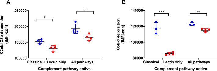
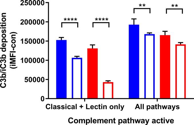
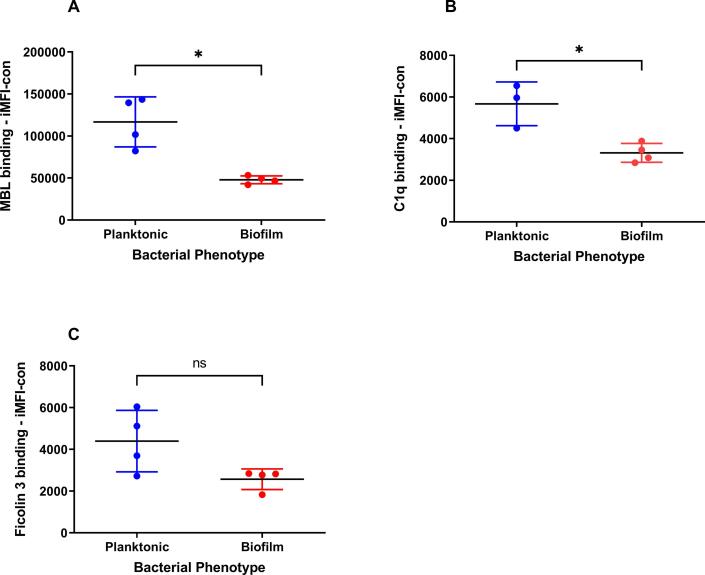
The development of new vaccines for TB needs to be underpinned by an understanding of both the molecular and cellular mechanisms of host-pathogen interactions and how the immune response can be modulated to achieve protection from disease. Complement orchestrates many aspects of the innate and adaptive immune responses. However, little is known about the contribution of the complement pathways during TB disease, particularly with respect to mycobacterial phenotype. Extracellular communities (biofilms) of M. tuberculosis are found in the acellular rim of granulomas, during disease, and these are likely to be present in post-primary TB episodes, in necrotic lesions. Our study aimed to determine which mycobacterial cell wall components were altered during biofilm growth and how these cell wall alterations modified the complement response. We have shown that M. tuberculosis biofilms modified their cell wall carbohydrates and elicited reduced classical and lectin pathway activation. Consistent with this finding was the reduction of C3b/iC3b deposition on biofilm cell wall carbohydrate extracts. Here, we have highlighted the role of cell wall carbohydrate alterations during biofilm growth of M. tuberculosis and subsequent modulation of complement activation.


 分享
分享
 求助内容:
求助内容: 应助结果提醒方式:
 扫码关注我们
