A Freilinger, K Kaserer, G Zettinig, P Pruidze, L F Reissig, T Rossmann, W J Weninger, S Meng
{"title":"甲状腺锥叶超声检查甲状腺锥体叶的超声检查","authors":"A Freilinger, K Kaserer, G Zettinig, P Pruidze, L F Reissig, T Rossmann, W J Weninger, S Meng","doi":"10.1007/s40618-022-01748-z","DOIUrl":null,"url":null,"abstract":"<p><strong>Purpose: </strong>The pyramidal lobe (PL) is an ancillary lobe of the thyroid gland that can be affected by the same pathologies as the rest of the gland. We aimed to assess the diagnostic performance of high-resolution sonography in the detection of the PL with verification by dissection and histological examination.</p><p><strong>Methods: </strong>In a prospective, cross-sectional mono-center study, 50 fresh, non-embalmed cadavers were included. Blinded ultrasound examination was performed to detect the PL by two investigators of different experience levels. If the PL was detected with ultrasound, dissection was performed to expose the PL and obtain a tissue sample. When no PL was detected with ultrasound, a tissue block of the anterior cervical region was excised. An endocrine pathologist microscopically examined all tissue samples and tissue blocks for the presence of thyroid parenchyma.</p><p><strong>Results: </strong>The prevalence of the PL was 80% [40/50; 95% CI (68.9%; 91.1%)]. Diagnostic performance for both examiners was: sensitivity (85.0%; 42.5%), specificity (50.0%; 60.0%), positive predictive value (87.2%; 81.0%), negative predictive value (45.5%; 21.0%) and accuracy (78.0%; 46.0%). Regression analysis demonstrated that neither thyroid parenchyma echogenicity, thyroid gland volume, age nor body size proved to be covariates in the accurate detection of a PL (p > .05).</p><p><strong>Conclusion: </strong>We report that high-resolution ultrasound is an adequate examination modality to detect the PL. Our findings indicate a higher prevalence than previously reported. Therefore, the PL may be regarded as a regular part of the thyroid gland. We also advocate a dedicated assessment of the PL in routine thyroid ultrasound.</p>","PeriodicalId":15651,"journal":{"name":"Journal of Endocrinological Investigation","volume":"45 6","pages":"1201-1208"},"PeriodicalIF":3.5000,"publicationDate":"2022-06-01","publicationTypes":"Journal Article","fieldsOfStudy":null,"isOpenAccess":false,"openAccessPdf":"https://www.ncbi.nlm.nih.gov/pmc/articles/PMC9098552/pdf/","citationCount":"2","resultStr":"{\"title\":\"Ultrasound for the detection of the pyramidal lobe of the thyroid gland.\",\"authors\":\"A Freilinger, K Kaserer, G Zettinig, P Pruidze, L F Reissig, T Rossmann, W J Weninger, S Meng\",\"doi\":\"10.1007/s40618-022-01748-z\",\"DOIUrl\":null,\"url\":null,\"abstract\":\"<p><strong>Purpose: </strong>The pyramidal lobe (PL) is an ancillary lobe of the thyroid gland that can be affected by the same pathologies as the rest of the gland. We aimed to assess the diagnostic performance of high-resolution sonography in the detection of the PL with verification by dissection and histological examination.</p><p><strong>Methods: </strong>In a prospective, cross-sectional mono-center study, 50 fresh, non-embalmed cadavers were included. Blinded ultrasound examination was performed to detect the PL by two investigators of different experience levels. If the PL was detected with ultrasound, dissection was performed to expose the PL and obtain a tissue sample. When no PL was detected with ultrasound, a tissue block of the anterior cervical region was excised. An endocrine pathologist microscopically examined all tissue samples and tissue blocks for the presence of thyroid parenchyma.</p><p><strong>Results: </strong>The prevalence of the PL was 80% [40/50; 95% CI (68.9%; 91.1%)]. Diagnostic performance for both examiners was: sensitivity (85.0%; 42.5%), specificity (50.0%; 60.0%), positive predictive value (87.2%; 81.0%), negative predictive value (45.5%; 21.0%) and accuracy (78.0%; 46.0%). Regression analysis demonstrated that neither thyroid parenchyma echogenicity, thyroid gland volume, age nor body size proved to be covariates in the accurate detection of a PL (p > .05).</p><p><strong>Conclusion: </strong>We report that high-resolution ultrasound is an adequate examination modality to detect the PL. Our findings indicate a higher prevalence than previously reported. Therefore, the PL may be regarded as a regular part of the thyroid gland. We also advocate a dedicated assessment of the PL in routine thyroid ultrasound.</p>\",\"PeriodicalId\":15651,\"journal\":{\"name\":\"Journal of Endocrinological Investigation\",\"volume\":\"45 6\",\"pages\":\"1201-1208\"},\"PeriodicalIF\":3.5000,\"publicationDate\":\"2022-06-01\",\"publicationTypes\":\"Journal Article\",\"fieldsOfStudy\":null,\"isOpenAccess\":false,\"openAccessPdf\":\"https://www.ncbi.nlm.nih.gov/pmc/articles/PMC9098552/pdf/\",\"citationCount\":\"2\",\"resultStr\":null,\"platform\":\"Semanticscholar\",\"paperid\":null,\"PeriodicalName\":\"Journal of Endocrinological Investigation\",\"FirstCategoryId\":\"3\",\"ListUrlMain\":\"https://doi.org/10.1007/s40618-022-01748-z\",\"RegionNum\":2,\"RegionCategory\":\"医学\",\"ArticlePicture\":[],\"TitleCN\":null,\"AbstractTextCN\":null,\"PMCID\":null,\"EPubDate\":\"2022/2/14 0:00:00\",\"PubModel\":\"Epub\",\"JCR\":\"Q2\",\"JCRName\":\"ENDOCRINOLOGY & METABOLISM\",\"Score\":null,\"Total\":0}","platform":"Semanticscholar","paperid":null,"PeriodicalName":"Journal of Endocrinological Investigation","FirstCategoryId":"3","ListUrlMain":"https://doi.org/10.1007/s40618-022-01748-z","RegionNum":2,"RegionCategory":"医学","ArticlePicture":[],"TitleCN":null,"AbstractTextCN":null,"PMCID":null,"EPubDate":"2022/2/14 0:00:00","PubModel":"Epub","JCR":"Q2","JCRName":"ENDOCRINOLOGY & METABOLISM","Score":null,"Total":0}
引用次数: 2
摘要
目的:锥体叶(PL)是甲状腺的辅助叶,可以受到与腺体其他部分相同的病理影响。我们的目的是通过解剖和组织学检查来评估高分辨率超声在PL检测中的诊断性能。方法:在一项前瞻性,横断面单中心研究中,包括50具新鲜,未防腐的尸体。由两名不同经验水平的调查员进行盲法超声检查以检测PL。如果用超声检测到PL,则进行解剖以暴露PL并获得组织样本。当超声未检测到PL时,切除宫颈前区组织块。内分泌病理学家在显微镜下检查了所有组织样本和组织块是否存在甲状腺实质。结果:PL患病率为80% [40/50];95% ci (68.9%;91.1%)]。两位检查者的诊断表现为:敏感性(85.0%);42.5%),特异性(50.0%;60.0%),阳性预测值为87.2%;81.0%),阴性预测值(45.5%;21.0%)和准确率(78.0%;46.0%)。回归分析表明,甲状腺实质回声、甲状腺体积、年龄和体型都不是准确检测PL的协变量(p > 0.05)。结论:我们报告,高分辨率超声是一种足够的检查方式来检测PL。我们的研究结果表明,比以前报道的患病率更高。因此,前甲状腺可视为甲状腺的一个规则部分。我们也提倡在常规甲状腺超声中对PL进行专门的评估。
Ultrasound for the detection of the pyramidal lobe of the thyroid gland.
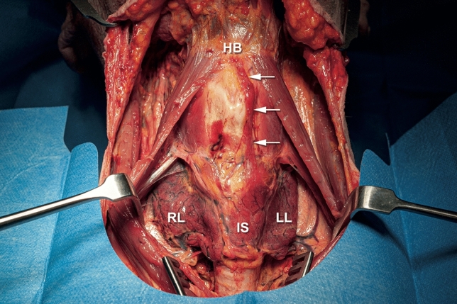
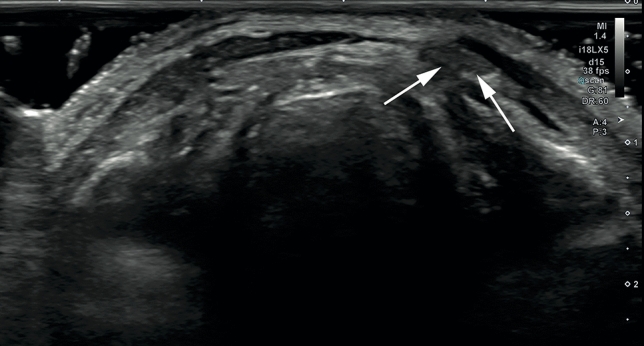
Purpose: The pyramidal lobe (PL) is an ancillary lobe of the thyroid gland that can be affected by the same pathologies as the rest of the gland. We aimed to assess the diagnostic performance of high-resolution sonography in the detection of the PL with verification by dissection and histological examination.
Methods: In a prospective, cross-sectional mono-center study, 50 fresh, non-embalmed cadavers were included. Blinded ultrasound examination was performed to detect the PL by two investigators of different experience levels. If the PL was detected with ultrasound, dissection was performed to expose the PL and obtain a tissue sample. When no PL was detected with ultrasound, a tissue block of the anterior cervical region was excised. An endocrine pathologist microscopically examined all tissue samples and tissue blocks for the presence of thyroid parenchyma.
Results: The prevalence of the PL was 80% [40/50; 95% CI (68.9%; 91.1%)]. Diagnostic performance for both examiners was: sensitivity (85.0%; 42.5%), specificity (50.0%; 60.0%), positive predictive value (87.2%; 81.0%), negative predictive value (45.5%; 21.0%) and accuracy (78.0%; 46.0%). Regression analysis demonstrated that neither thyroid parenchyma echogenicity, thyroid gland volume, age nor body size proved to be covariates in the accurate detection of a PL (p > .05).
Conclusion: We report that high-resolution ultrasound is an adequate examination modality to detect the PL. Our findings indicate a higher prevalence than previously reported. Therefore, the PL may be regarded as a regular part of the thyroid gland. We also advocate a dedicated assessment of the PL in routine thyroid ultrasound.
期刊介绍:
The Journal of Endocrinological Investigation is a well-established, e-only endocrine journal founded 36 years ago in 1978. It is the official journal of the Italian Society of Endocrinology (SIE), established in 1964. Other Italian societies in the endocrinology and metabolism field are affiliated to the journal: Italian Society of Andrology and Sexual Medicine, Italian Society of Obesity, Italian Society of Pediatric Endocrinology and Diabetology, Clinical Endocrinologists’ Association, Thyroid Association, Endocrine Surgical Units Association, Italian Society of Pharmacology.


 分享
分享
 求助内容:
求助内容: 应助结果提醒方式:
 扫码关注我们
