{"title":"始祖舌藻低聚糖转移酶第三副c端球状结构域的晶体结构","authors":"Shunsuke Matsumoto, Atsushi Shimada, Daisuke Kohda","doi":"10.1186/1472-6807-13-11","DOIUrl":null,"url":null,"abstract":"<p>Protein N-glycosylation occurs in the three domains of life. Oligosaccharyltransferase (OST) transfers an oligosaccharide chain to the asparagine residue in the N-glycosylation sequons. The catalytic subunits of the OST enzyme are STT3 in eukaryotes, AglB in archaea and PglB in eubacteria. The genome of a hyperthermophilic archaeon, <i>Archaeoglobus fulgidus</i>, encodes three paralogous AglB proteins. We previously solved the crystal structures of the C-terminal globular domains of two paralogs, AglB-S<i>hort</i> 1 and AglB-S<i>hort</i> 2.</p><p>We determined the crystal structure of the C-terminal globular domain of the third AglB paralog, AglB-L<i>ong</i>, at 1.9?? resolutions. The crystallization of the fusion protein with maltose binding protein (MBP) afforded high quality protein crystals. Two MBP-AglB-L molecules formed a swapped dimer in the crystal. Since the fusion protein behaved as a monomer upon gel filtration, we reconstituted the monomer structure from the swapped dimer by exchanging the swapped segments. The C-terminal domain of <i>A. fulgidus</i> AglB-L includes a structural unit common to AglB-S1 and AglB-S2. This structural unit contains the evolutionally conserved WWDYG and DK motifs. The present structure revealed that <i>A. fulgidus</i> AglB-L contained a variant type of the DK motif with a short insertion, and confirmed that the second signature residue, Lys, of the DK motif participates in the formation of a pocket that binds to the serine and threonine residues at the +2 position of the N-glycosylation sequon.</p><p>The structure of <i>A. fulgidus</i> AglB-L, together with the two previously solved structures of AglB-S1 and AglB-S2, provides a complete overview of the three AglB paralogs encoded in the <i>A. fulgidus</i> genome. All three AglBs contain a variant type of the DK motif. This finding supports a previously proposed rule: The STT3/AglB/PglB paralogs in one organism always contain the same type of Ser/Thr-binding pocket. The present structure will be useful as a search model for molecular replacement in the structural determination of the full-length <i>A. fulgidus</i> AglB-L.</p>","PeriodicalId":498,"journal":{"name":"BMC Structural Biology","volume":"13 1","pages":""},"PeriodicalIF":2.2220,"publicationDate":"2013-07-01","publicationTypes":"Journal Article","fieldsOfStudy":null,"isOpenAccess":false,"openAccessPdf":"https://sci-hub-pdf.com/10.1186/1472-6807-13-11","citationCount":"28","resultStr":"{\"title\":\"Crystal structure of the C-terminal globular domain of the third paralog of the Archaeoglobus fulgidus oligosaccharyltransferases\",\"authors\":\"Shunsuke Matsumoto, Atsushi Shimada, Daisuke Kohda\",\"doi\":\"10.1186/1472-6807-13-11\",\"DOIUrl\":null,\"url\":null,\"abstract\":\"<p>Protein N-glycosylation occurs in the three domains of life. Oligosaccharyltransferase (OST) transfers an oligosaccharide chain to the asparagine residue in the N-glycosylation sequons. The catalytic subunits of the OST enzyme are STT3 in eukaryotes, AglB in archaea and PglB in eubacteria. The genome of a hyperthermophilic archaeon, <i>Archaeoglobus fulgidus</i>, encodes three paralogous AglB proteins. We previously solved the crystal structures of the C-terminal globular domains of two paralogs, AglB-S<i>hort</i> 1 and AglB-S<i>hort</i> 2.</p><p>We determined the crystal structure of the C-terminal globular domain of the third AglB paralog, AglB-L<i>ong</i>, at 1.9?? resolutions. The crystallization of the fusion protein with maltose binding protein (MBP) afforded high quality protein crystals. Two MBP-AglB-L molecules formed a swapped dimer in the crystal. Since the fusion protein behaved as a monomer upon gel filtration, we reconstituted the monomer structure from the swapped dimer by exchanging the swapped segments. The C-terminal domain of <i>A. fulgidus</i> AglB-L includes a structural unit common to AglB-S1 and AglB-S2. This structural unit contains the evolutionally conserved WWDYG and DK motifs. The present structure revealed that <i>A. fulgidus</i> AglB-L contained a variant type of the DK motif with a short insertion, and confirmed that the second signature residue, Lys, of the DK motif participates in the formation of a pocket that binds to the serine and threonine residues at the +2 position of the N-glycosylation sequon.</p><p>The structure of <i>A. fulgidus</i> AglB-L, together with the two previously solved structures of AglB-S1 and AglB-S2, provides a complete overview of the three AglB paralogs encoded in the <i>A. fulgidus</i> genome. All three AglBs contain a variant type of the DK motif. This finding supports a previously proposed rule: The STT3/AglB/PglB paralogs in one organism always contain the same type of Ser/Thr-binding pocket. The present structure will be useful as a search model for molecular replacement in the structural determination of the full-length <i>A. fulgidus</i> AglB-L.</p>\",\"PeriodicalId\":498,\"journal\":{\"name\":\"BMC Structural Biology\",\"volume\":\"13 1\",\"pages\":\"\"},\"PeriodicalIF\":2.2220,\"publicationDate\":\"2013-07-01\",\"publicationTypes\":\"Journal Article\",\"fieldsOfStudy\":null,\"isOpenAccess\":false,\"openAccessPdf\":\"https://sci-hub-pdf.com/10.1186/1472-6807-13-11\",\"citationCount\":\"28\",\"resultStr\":null,\"platform\":\"Semanticscholar\",\"paperid\":null,\"PeriodicalName\":\"BMC Structural Biology\",\"FirstCategoryId\":\"1085\",\"ListUrlMain\":\"https://link.springer.com/article/10.1186/1472-6807-13-11\",\"RegionNum\":0,\"RegionCategory\":null,\"ArticlePicture\":[],\"TitleCN\":null,\"AbstractTextCN\":null,\"PMCID\":null,\"EPubDate\":\"\",\"PubModel\":\"\",\"JCR\":\"Q3\",\"JCRName\":\"Biochemistry, Genetics and Molecular Biology\",\"Score\":null,\"Total\":0}","platform":"Semanticscholar","paperid":null,"PeriodicalName":"BMC Structural Biology","FirstCategoryId":"1085","ListUrlMain":"https://link.springer.com/article/10.1186/1472-6807-13-11","RegionNum":0,"RegionCategory":null,"ArticlePicture":[],"TitleCN":null,"AbstractTextCN":null,"PMCID":null,"EPubDate":"","PubModel":"","JCR":"Q3","JCRName":"Biochemistry, Genetics and Molecular Biology","Score":null,"Total":0}
Crystal structure of the C-terminal globular domain of the third paralog of the Archaeoglobus fulgidus oligosaccharyltransferases
Protein N-glycosylation occurs in the three domains of life. Oligosaccharyltransferase (OST) transfers an oligosaccharide chain to the asparagine residue in the N-glycosylation sequons. The catalytic subunits of the OST enzyme are STT3 in eukaryotes, AglB in archaea and PglB in eubacteria. The genome of a hyperthermophilic archaeon, Archaeoglobus fulgidus, encodes three paralogous AglB proteins. We previously solved the crystal structures of the C-terminal globular domains of two paralogs, AglB-Short 1 and AglB-Short 2.
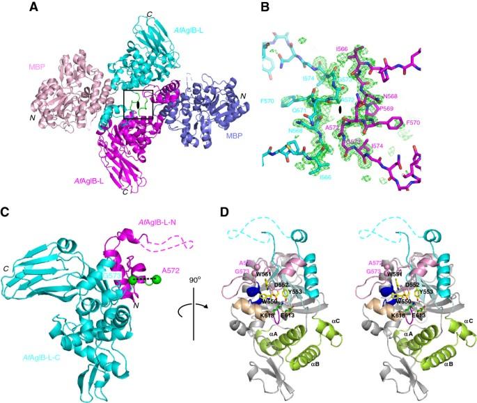
We determined the crystal structure of the C-terminal globular domain of the third AglB paralog, AglB-Long, at 1.9?? resolutions. The crystallization of the fusion protein with maltose binding protein (MBP) afforded high quality protein crystals. Two MBP-AglB-L molecules formed a swapped dimer in the crystal. Since the fusion protein behaved as a monomer upon gel filtration, we reconstituted the monomer structure from the swapped dimer by exchanging the swapped segments. The C-terminal domain of A. fulgidus AglB-L includes a structural unit common to AglB-S1 and AglB-S2. This structural unit contains the evolutionally conserved WWDYG and DK motifs. The present structure revealed that A. fulgidus AglB-L contained a variant type of the DK motif with a short insertion, and confirmed that the second signature residue, Lys, of the DK motif participates in the formation of a pocket that binds to the serine and threonine residues at the +2 position of the N-glycosylation sequon.
The structure of A. fulgidus AglB-L, together with the two previously solved structures of AglB-S1 and AglB-S2, provides a complete overview of the three AglB paralogs encoded in the A. fulgidus genome. All three AglBs contain a variant type of the DK motif. This finding supports a previously proposed rule: The STT3/AglB/PglB paralogs in one organism always contain the same type of Ser/Thr-binding pocket. The present structure will be useful as a search model for molecular replacement in the structural determination of the full-length A. fulgidus AglB-L.
期刊介绍:
BMC Structural Biology is an open access, peer-reviewed journal that considers articles on investigations into the structure of biological macromolecules, including solving structures, structural and functional analyses, and computational modeling.


 分享
分享
 求助内容:
求助内容: 应助结果提醒方式:
 扫码关注我们
