{"title":"杜氏肌营养不良大鼠舌肌大舌失音及不太严重的营养不良改变。","authors":"Keitaro Yamanouchi, Yukie Tanaka, Masanari Ikeda, Shizuka Kato, Ryosuke Okino, Hiroki Nishi, Fumihiko Hakuno, Shin-Ichiro Takahashi, James Chambers, Takashi Matsuwaki, Kazuyuki Uchida","doi":"10.1186/s13395-022-00307-7","DOIUrl":null,"url":null,"abstract":"<p><strong>Background: </strong>Duchenne muscular dystrophy (DMD) is an X-linked muscle disease caused by a complete lack of dystrophin, which stabilizes the plasma membrane of myofibers. The orofacial function is affected in an advanced stage of DMD and this often leads to an eating disorder such as dysphagia. Dysphagia is caused by multiple etiologies including decreased mastication and swallowing. Therefore, preventing the functional declines of mastication and swallowing in DMD is important to improve the patient's quality of life. In the present study, using a rat model of DMD we generated previously, we performed analyses on the masseter and tongue muscles, both are required for proper eating function.</p><p><strong>Methods: </strong>Age-related changes of the masseter and tongue muscle of DMD rats were analyzed morphometrically, histologically, and immunohistochemically. Also, transcription of cellular senescent markers, and utrophin (Utrn), a functional analog of dystrophin, was examined.</p><p><strong>Results: </strong>The masseter muscle of DMD rats showed progressive dystrophic changes as observed in their hindlimb muscle, accompanied by increased transcription of p16 and p19. On the other hand, the tongue of DMD rats showed macroglossia due to hypertrophy of myofibers with less dystrophic changes. Proliferative activity was preserved in the satellite cells from the tongue muscle but was perturbed severely in those from the masseter muscle. While Utrn transcription was increased in the masseter muscle of DMD rats compared to WT rats, probably due to a compensatory mechanism, its level in the tongue muscle was comparable between WT and DMD rats and was similar to that in the masseter muscle of DMD rats.</p><p><strong>Conclusions: </strong>Muscular dystrophy is less advanced in the tongue muscle compared to the masseter muscle in the DMD rat.</p>","PeriodicalId":21747,"journal":{"name":"Skeletal Muscle","volume":" ","pages":"24"},"PeriodicalIF":4.4000,"publicationDate":"2022-10-19","publicationTypes":"Journal Article","fieldsOfStudy":null,"isOpenAccess":false,"openAccessPdf":"https://www.ncbi.nlm.nih.gov/pmc/articles/PMC9580129/pdf/","citationCount":"2","resultStr":"{\"title\":\"Macroglossia and less advanced dystrophic change in the tongue muscle of the Duchenne muscular dystrophy rat.\",\"authors\":\"Keitaro Yamanouchi, Yukie Tanaka, Masanari Ikeda, Shizuka Kato, Ryosuke Okino, Hiroki Nishi, Fumihiko Hakuno, Shin-Ichiro Takahashi, James Chambers, Takashi Matsuwaki, Kazuyuki Uchida\",\"doi\":\"10.1186/s13395-022-00307-7\",\"DOIUrl\":null,\"url\":null,\"abstract\":\"<p><strong>Background: </strong>Duchenne muscular dystrophy (DMD) is an X-linked muscle disease caused by a complete lack of dystrophin, which stabilizes the plasma membrane of myofibers. The orofacial function is affected in an advanced stage of DMD and this often leads to an eating disorder such as dysphagia. Dysphagia is caused by multiple etiologies including decreased mastication and swallowing. Therefore, preventing the functional declines of mastication and swallowing in DMD is important to improve the patient's quality of life. In the present study, using a rat model of DMD we generated previously, we performed analyses on the masseter and tongue muscles, both are required for proper eating function.</p><p><strong>Methods: </strong>Age-related changes of the masseter and tongue muscle of DMD rats were analyzed morphometrically, histologically, and immunohistochemically. Also, transcription of cellular senescent markers, and utrophin (Utrn), a functional analog of dystrophin, was examined.</p><p><strong>Results: </strong>The masseter muscle of DMD rats showed progressive dystrophic changes as observed in their hindlimb muscle, accompanied by increased transcription of p16 and p19. On the other hand, the tongue of DMD rats showed macroglossia due to hypertrophy of myofibers with less dystrophic changes. Proliferative activity was preserved in the satellite cells from the tongue muscle but was perturbed severely in those from the masseter muscle. While Utrn transcription was increased in the masseter muscle of DMD rats compared to WT rats, probably due to a compensatory mechanism, its level in the tongue muscle was comparable between WT and DMD rats and was similar to that in the masseter muscle of DMD rats.</p><p><strong>Conclusions: </strong>Muscular dystrophy is less advanced in the tongue muscle compared to the masseter muscle in the DMD rat.</p>\",\"PeriodicalId\":21747,\"journal\":{\"name\":\"Skeletal Muscle\",\"volume\":\" \",\"pages\":\"24\"},\"PeriodicalIF\":4.4000,\"publicationDate\":\"2022-10-19\",\"publicationTypes\":\"Journal Article\",\"fieldsOfStudy\":null,\"isOpenAccess\":false,\"openAccessPdf\":\"https://www.ncbi.nlm.nih.gov/pmc/articles/PMC9580129/pdf/\",\"citationCount\":\"2\",\"resultStr\":null,\"platform\":\"Semanticscholar\",\"paperid\":null,\"PeriodicalName\":\"Skeletal Muscle\",\"FirstCategoryId\":\"3\",\"ListUrlMain\":\"https://doi.org/10.1186/s13395-022-00307-7\",\"RegionNum\":2,\"RegionCategory\":\"医学\",\"ArticlePicture\":[],\"TitleCN\":null,\"AbstractTextCN\":null,\"PMCID\":null,\"EPubDate\":\"\",\"PubModel\":\"\",\"JCR\":\"Q2\",\"JCRName\":\"CELL BIOLOGY\",\"Score\":null,\"Total\":0}","platform":"Semanticscholar","paperid":null,"PeriodicalName":"Skeletal Muscle","FirstCategoryId":"3","ListUrlMain":"https://doi.org/10.1186/s13395-022-00307-7","RegionNum":2,"RegionCategory":"医学","ArticlePicture":[],"TitleCN":null,"AbstractTextCN":null,"PMCID":null,"EPubDate":"","PubModel":"","JCR":"Q2","JCRName":"CELL BIOLOGY","Score":null,"Total":0}
Macroglossia and less advanced dystrophic change in the tongue muscle of the Duchenne muscular dystrophy rat.
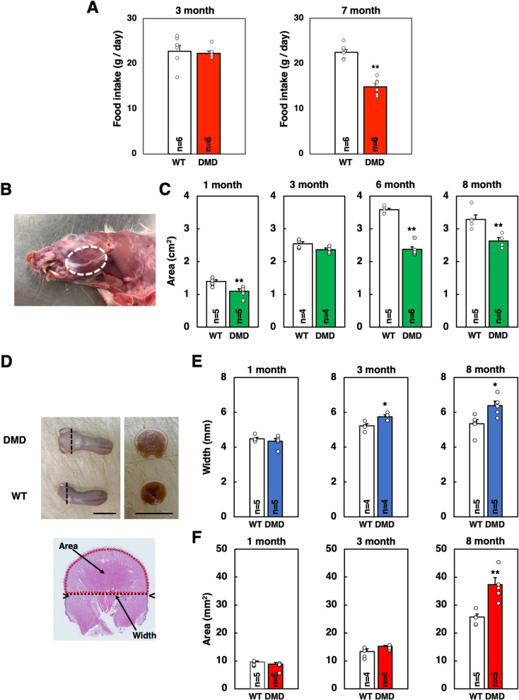
Background: Duchenne muscular dystrophy (DMD) is an X-linked muscle disease caused by a complete lack of dystrophin, which stabilizes the plasma membrane of myofibers. The orofacial function is affected in an advanced stage of DMD and this often leads to an eating disorder such as dysphagia. Dysphagia is caused by multiple etiologies including decreased mastication and swallowing. Therefore, preventing the functional declines of mastication and swallowing in DMD is important to improve the patient's quality of life. In the present study, using a rat model of DMD we generated previously, we performed analyses on the masseter and tongue muscles, both are required for proper eating function.
Methods: Age-related changes of the masseter and tongue muscle of DMD rats were analyzed morphometrically, histologically, and immunohistochemically. Also, transcription of cellular senescent markers, and utrophin (Utrn), a functional analog of dystrophin, was examined.
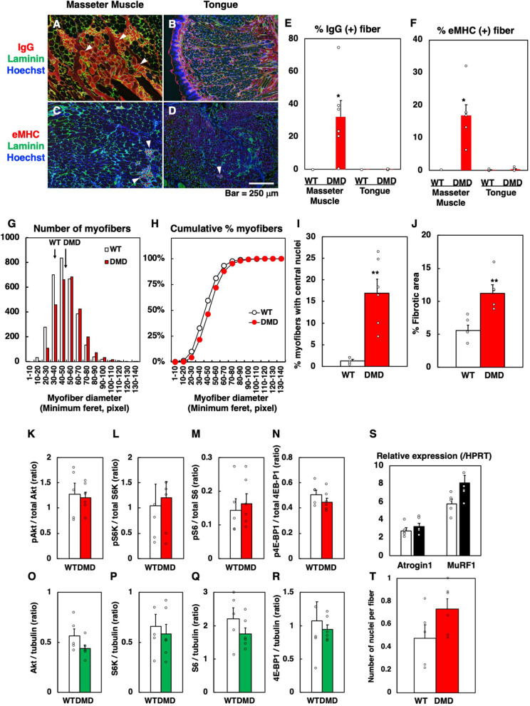
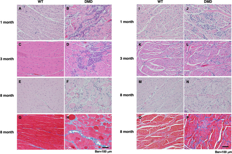
Results: The masseter muscle of DMD rats showed progressive dystrophic changes as observed in their hindlimb muscle, accompanied by increased transcription of p16 and p19. On the other hand, the tongue of DMD rats showed macroglossia due to hypertrophy of myofibers with less dystrophic changes. Proliferative activity was preserved in the satellite cells from the tongue muscle but was perturbed severely in those from the masseter muscle. While Utrn transcription was increased in the masseter muscle of DMD rats compared to WT rats, probably due to a compensatory mechanism, its level in the tongue muscle was comparable between WT and DMD rats and was similar to that in the masseter muscle of DMD rats.
Conclusions: Muscular dystrophy is less advanced in the tongue muscle compared to the masseter muscle in the DMD rat.
期刊介绍:
The only open access journal in its field, Skeletal Muscle publishes novel, cutting-edge research and technological advancements that investigate the molecular mechanisms underlying the biology of skeletal muscle. Reflecting the breadth of research in this area, the journal welcomes manuscripts about the development, metabolism, the regulation of mass and function, aging, degeneration, dystrophy and regeneration of skeletal muscle, with an emphasis on understanding adult skeletal muscle, its maintenance, and its interactions with non-muscle cell types and regulatory modulators.
Main areas of interest include:
-differentiation of skeletal muscle-
atrophy and hypertrophy of skeletal muscle-
aging of skeletal muscle-
regeneration and degeneration of skeletal muscle-
biology of satellite and satellite-like cells-
dystrophic degeneration of skeletal muscle-
energy and glucose homeostasis in skeletal muscle-
non-dystrophic genetic diseases of skeletal muscle, such as Spinal Muscular Atrophy and myopathies-
maintenance of neuromuscular junctions-
roles of ryanodine receptors and calcium signaling in skeletal muscle-
roles of nuclear receptors in skeletal muscle-
roles of GPCRs and GPCR signaling in skeletal muscle-
other relevant aspects of skeletal muscle biology.
In addition, articles on translational clinical studies that address molecular and cellular mechanisms of skeletal muscle will be published. Case reports are also encouraged for submission.
Skeletal Muscle reflects the breadth of research on skeletal muscle and bridges gaps between diverse areas of science for example cardiac cell biology and neurobiology, which share common features with respect to cell differentiation, excitatory membranes, cell-cell communication, and maintenance. Suitable articles are model and mechanism-driven, and apply statistical principles where appropriate; purely descriptive studies are of lesser interest.


 分享
分享
 求助内容:
求助内容: 应助结果提醒方式:
 扫码关注我们
