Krystian Truszkiewicz, Piotr Macek, Małgorzata Poręba, Rafał Poręba, Paweł Gać
{"title":"超声心动图评价胸心比值作为左心室肥厚的潜在标志。","authors":"Krystian Truszkiewicz, Piotr Macek, Małgorzata Poręba, Rafał Poręba, Paweł Gać","doi":"10.1155/2022/4931945","DOIUrl":null,"url":null,"abstract":"<p><p>The aim of the study was to verify the usefulness of the radiological cardiothoracic ratio as a potential marker of left ventricular hypertrophy assessed by echocardiography. The study included 96 patients (mean age: 49.52 ± 9.64 years). Chest radiograph in the PA projection and echocardiography were performed. In CR the measurement of the cardiothoracic ratio (CTR) was performed. Assuming CTR > 0.50, heart silhouette enlargement was diagnosed. In echocardiography, four types of left ventricular geometry were assessed: normal geometry (NG), concentric remodeling (CR), concentric hypertrophy (CH), and eccentric hypertrophy (EH). It was shown that patients with an enlarged heart silhouette were characterized by a significantly more frequent occurrence of left ventricular hypertrophy (LVH) on echocardiography than patients with a nonenlarged heart silhouette. In the subgroup of patients with LVH compared to the subgroup of patients with normal left ventricular geometry, CTR values are statistically significantly higher, and heart silhouette enlargement is significantly more frequent. The criterion \"CTR > 0.49\" estimates LVH with a sensitivity of 93.3% and specificity of 82.7%, which translates into a high accuracy of 84.4%. By analyzing the prediction of left ventricular geometry types, high accuracy of CH prediction was obtained using the \"CTR > 0.49\" criterion of 80.2% (with a high sensitivity of 84.0% and a satisfactory specificity of 60.0%) and a high accuracy of EH prediction using the \"CTR > 0.52\" criterion of 71.9% (with high sensitivity 80.5% and low specificity 36.8%), as well as low CR prediction accuracy of only 57.3% (with low sensitivity 36.7%, even if high specificity 78.7%). In summary, the radiological cardiothoracic ratio may be a moderate marker of left ventricular hypertrophy assessed according to standard echocardiographic criteria, provided that its cut-off point is standardized in each population of subjects.</p>","PeriodicalId":51864,"journal":{"name":"Radiology Research and Practice","volume":" ","pages":"4931945"},"PeriodicalIF":1.5000,"publicationDate":"2022-06-15","publicationTypes":"Journal Article","fieldsOfStudy":null,"isOpenAccess":false,"openAccessPdf":"https://www.ncbi.nlm.nih.gov/pmc/articles/PMC9217623/pdf/","citationCount":"2","resultStr":"{\"title\":\"Radiological Cardiothoracic Ratio as a Potential Marker of Left Ventricular Hypertrophy Assessed by Echocardiography.\",\"authors\":\"Krystian Truszkiewicz, Piotr Macek, Małgorzata Poręba, Rafał Poręba, Paweł Gać\",\"doi\":\"10.1155/2022/4931945\",\"DOIUrl\":null,\"url\":null,\"abstract\":\"<p><p>The aim of the study was to verify the usefulness of the radiological cardiothoracic ratio as a potential marker of left ventricular hypertrophy assessed by echocardiography. The study included 96 patients (mean age: 49.52 ± 9.64 years). Chest radiograph in the PA projection and echocardiography were performed. In CR the measurement of the cardiothoracic ratio (CTR) was performed. Assuming CTR > 0.50, heart silhouette enlargement was diagnosed. In echocardiography, four types of left ventricular geometry were assessed: normal geometry (NG), concentric remodeling (CR), concentric hypertrophy (CH), and eccentric hypertrophy (EH). It was shown that patients with an enlarged heart silhouette were characterized by a significantly more frequent occurrence of left ventricular hypertrophy (LVH) on echocardiography than patients with a nonenlarged heart silhouette. In the subgroup of patients with LVH compared to the subgroup of patients with normal left ventricular geometry, CTR values are statistically significantly higher, and heart silhouette enlargement is significantly more frequent. The criterion \\\"CTR > 0.49\\\" estimates LVH with a sensitivity of 93.3% and specificity of 82.7%, which translates into a high accuracy of 84.4%. By analyzing the prediction of left ventricular geometry types, high accuracy of CH prediction was obtained using the \\\"CTR > 0.49\\\" criterion of 80.2% (with a high sensitivity of 84.0% and a satisfactory specificity of 60.0%) and a high accuracy of EH prediction using the \\\"CTR > 0.52\\\" criterion of 71.9% (with high sensitivity 80.5% and low specificity 36.8%), as well as low CR prediction accuracy of only 57.3% (with low sensitivity 36.7%, even if high specificity 78.7%). In summary, the radiological cardiothoracic ratio may be a moderate marker of left ventricular hypertrophy assessed according to standard echocardiographic criteria, provided that its cut-off point is standardized in each population of subjects.</p>\",\"PeriodicalId\":51864,\"journal\":{\"name\":\"Radiology Research and Practice\",\"volume\":\" \",\"pages\":\"4931945\"},\"PeriodicalIF\":1.5000,\"publicationDate\":\"2022-06-15\",\"publicationTypes\":\"Journal Article\",\"fieldsOfStudy\":null,\"isOpenAccess\":false,\"openAccessPdf\":\"https://www.ncbi.nlm.nih.gov/pmc/articles/PMC9217623/pdf/\",\"citationCount\":\"2\",\"resultStr\":null,\"platform\":\"Semanticscholar\",\"paperid\":null,\"PeriodicalName\":\"Radiology Research and Practice\",\"FirstCategoryId\":\"1085\",\"ListUrlMain\":\"https://doi.org/10.1155/2022/4931945\",\"RegionNum\":0,\"RegionCategory\":null,\"ArticlePicture\":[],\"TitleCN\":null,\"AbstractTextCN\":null,\"PMCID\":null,\"EPubDate\":\"2022/1/1 0:00:00\",\"PubModel\":\"eCollection\",\"JCR\":\"Q2\",\"JCRName\":\"RADIOLOGY, NUCLEAR MEDICINE & MEDICAL IMAGING\",\"Score\":null,\"Total\":0}","platform":"Semanticscholar","paperid":null,"PeriodicalName":"Radiology Research and Practice","FirstCategoryId":"1085","ListUrlMain":"https://doi.org/10.1155/2022/4931945","RegionNum":0,"RegionCategory":null,"ArticlePicture":[],"TitleCN":null,"AbstractTextCN":null,"PMCID":null,"EPubDate":"2022/1/1 0:00:00","PubModel":"eCollection","JCR":"Q2","JCRName":"RADIOLOGY, NUCLEAR MEDICINE & MEDICAL IMAGING","Score":null,"Total":0}
Radiological Cardiothoracic Ratio as a Potential Marker of Left Ventricular Hypertrophy Assessed by Echocardiography.
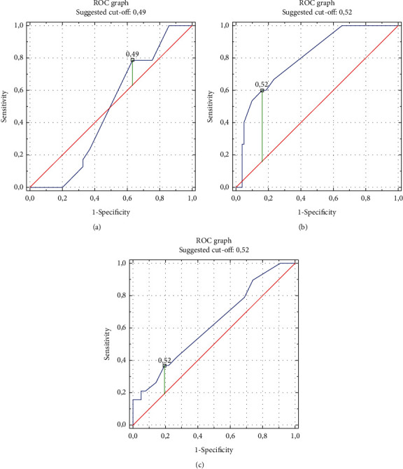
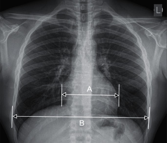
The aim of the study was to verify the usefulness of the radiological cardiothoracic ratio as a potential marker of left ventricular hypertrophy assessed by echocardiography. The study included 96 patients (mean age: 49.52 ± 9.64 years). Chest radiograph in the PA projection and echocardiography were performed. In CR the measurement of the cardiothoracic ratio (CTR) was performed. Assuming CTR > 0.50, heart silhouette enlargement was diagnosed. In echocardiography, four types of left ventricular geometry were assessed: normal geometry (NG), concentric remodeling (CR), concentric hypertrophy (CH), and eccentric hypertrophy (EH). It was shown that patients with an enlarged heart silhouette were characterized by a significantly more frequent occurrence of left ventricular hypertrophy (LVH) on echocardiography than patients with a nonenlarged heart silhouette. In the subgroup of patients with LVH compared to the subgroup of patients with normal left ventricular geometry, CTR values are statistically significantly higher, and heart silhouette enlargement is significantly more frequent. The criterion "CTR > 0.49" estimates LVH with a sensitivity of 93.3% and specificity of 82.7%, which translates into a high accuracy of 84.4%. By analyzing the prediction of left ventricular geometry types, high accuracy of CH prediction was obtained using the "CTR > 0.49" criterion of 80.2% (with a high sensitivity of 84.0% and a satisfactory specificity of 60.0%) and a high accuracy of EH prediction using the "CTR > 0.52" criterion of 71.9% (with high sensitivity 80.5% and low specificity 36.8%), as well as low CR prediction accuracy of only 57.3% (with low sensitivity 36.7%, even if high specificity 78.7%). In summary, the radiological cardiothoracic ratio may be a moderate marker of left ventricular hypertrophy assessed according to standard echocardiographic criteria, provided that its cut-off point is standardized in each population of subjects.
期刊介绍:
Radiology Research and Practice is a peer-reviewed, Open Access journal that publishes articles on all areas of medical imaging. The journal promotes evidence-based radiology practice though the publication of original research, reviews, and clinical studies for a multidisciplinary audience. Radiology Research and Practice is archived in Portico, which provides permanent archiving for electronic scholarly journals, as well as via the LOCKSS initiative. It operates a fully open access publishing model which allows open global access to its published content. This model is supported through Article Processing Charges. For more information on Article Processing charges in gen


 分享
分享
 求助内容:
求助内容: 应助结果提醒方式:
 扫码关注我们
