Mirna Radovic, Lidia Gavic, Daniel Jerkovic, Davor Zeljezic, Jasna Puizina, Ivan Srzentic, Ema Puizina Mladinic, Antonija Tadin
{"title":"牙种植体对牙龈上皮细胞基因毒性作用的临床前瞻性评估。","authors":"Mirna Radovic, Lidia Gavic, Daniel Jerkovic, Davor Zeljezic, Jasna Puizina, Ivan Srzentic, Ema Puizina Mladinic, Antonija Tadin","doi":"10.15644/asc56/3/1","DOIUrl":null,"url":null,"abstract":"<p><strong>Objectives: </strong>Although titanium-based implants are considered bioinert, it has been found that they are subject to corrosion and wear. This study aimed to evaluate the cytotoxic and genotoxic potential of two implant systems in gingival epithelial cells.</p><p><strong>Material and methods: </strong>Gingival swabs were taken three times from 91 subjects. The first swab was taken before dental implant placement, the second swab 90 days after dental implant placement and the third swab 21 days following the healing abutment placement. DNA damage was analyzed using the micronucleus test. Tested dental implants with corresponding healing abutments were Ankylos and Dentium SuperLine.</p><p><strong>Results: </strong>Of all scored forms of cytogenetic damage in gingival cells of individuals after implementation of tested dental implant systems, only an increase in the number of binucleated cells (P ≤ 0.001) was significant in contrast to control values for both tested implant systems, 90 days after dental implant placement and 21 days following the healing abutment placement.</p><p><strong>Conclusion: </strong>It may be concluded that there are no titanium-based implant dependent cytogenetic damage in gingival epithelial cells. A slight increase in cytogenetic damage has been observed but it is of no biological relevance and might be associated with healing abutment induced effect.</p>","PeriodicalId":7154,"journal":{"name":"Acta Stomatologica Croatica","volume":"56 3","pages":"222-234"},"PeriodicalIF":1.8000,"publicationDate":"2022-09-01","publicationTypes":"Journal Article","fieldsOfStudy":null,"isOpenAccess":false,"openAccessPdf":"https://ftp.ncbi.nlm.nih.gov/pub/pmc/oa_pdf/c4/e2/ASC_56(3)_222-234.PMC9635501.pdf","citationCount":"2","resultStr":"{\"title\":\"Clinical Prospective Assessment of Genotoxic Effects of Dental Implants in Gingival Epithelial Cells.\",\"authors\":\"Mirna Radovic, Lidia Gavic, Daniel Jerkovic, Davor Zeljezic, Jasna Puizina, Ivan Srzentic, Ema Puizina Mladinic, Antonija Tadin\",\"doi\":\"10.15644/asc56/3/1\",\"DOIUrl\":null,\"url\":null,\"abstract\":\"<p><strong>Objectives: </strong>Although titanium-based implants are considered bioinert, it has been found that they are subject to corrosion and wear. This study aimed to evaluate the cytotoxic and genotoxic potential of two implant systems in gingival epithelial cells.</p><p><strong>Material and methods: </strong>Gingival swabs were taken three times from 91 subjects. The first swab was taken before dental implant placement, the second swab 90 days after dental implant placement and the third swab 21 days following the healing abutment placement. DNA damage was analyzed using the micronucleus test. Tested dental implants with corresponding healing abutments were Ankylos and Dentium SuperLine.</p><p><strong>Results: </strong>Of all scored forms of cytogenetic damage in gingival cells of individuals after implementation of tested dental implant systems, only an increase in the number of binucleated cells (P ≤ 0.001) was significant in contrast to control values for both tested implant systems, 90 days after dental implant placement and 21 days following the healing abutment placement.</p><p><strong>Conclusion: </strong>It may be concluded that there are no titanium-based implant dependent cytogenetic damage in gingival epithelial cells. A slight increase in cytogenetic damage has been observed but it is of no biological relevance and might be associated with healing abutment induced effect.</p>\",\"PeriodicalId\":7154,\"journal\":{\"name\":\"Acta Stomatologica Croatica\",\"volume\":\"56 3\",\"pages\":\"222-234\"},\"PeriodicalIF\":1.8000,\"publicationDate\":\"2022-09-01\",\"publicationTypes\":\"Journal Article\",\"fieldsOfStudy\":null,\"isOpenAccess\":false,\"openAccessPdf\":\"https://ftp.ncbi.nlm.nih.gov/pub/pmc/oa_pdf/c4/e2/ASC_56(3)_222-234.PMC9635501.pdf\",\"citationCount\":\"2\",\"resultStr\":null,\"platform\":\"Semanticscholar\",\"paperid\":null,\"PeriodicalName\":\"Acta Stomatologica Croatica\",\"FirstCategoryId\":\"1085\",\"ListUrlMain\":\"https://doi.org/10.15644/asc56/3/1\",\"RegionNum\":0,\"RegionCategory\":null,\"ArticlePicture\":[],\"TitleCN\":null,\"AbstractTextCN\":null,\"PMCID\":null,\"EPubDate\":\"\",\"PubModel\":\"\",\"JCR\":\"Q3\",\"JCRName\":\"DENTISTRY, ORAL SURGERY & MEDICINE\",\"Score\":null,\"Total\":0}","platform":"Semanticscholar","paperid":null,"PeriodicalName":"Acta Stomatologica Croatica","FirstCategoryId":"1085","ListUrlMain":"https://doi.org/10.15644/asc56/3/1","RegionNum":0,"RegionCategory":null,"ArticlePicture":[],"TitleCN":null,"AbstractTextCN":null,"PMCID":null,"EPubDate":"","PubModel":"","JCR":"Q3","JCRName":"DENTISTRY, ORAL SURGERY & MEDICINE","Score":null,"Total":0}
Clinical Prospective Assessment of Genotoxic Effects of Dental Implants in Gingival Epithelial Cells.
Objectives: Although titanium-based implants are considered bioinert, it has been found that they are subject to corrosion and wear. This study aimed to evaluate the cytotoxic and genotoxic potential of two implant systems in gingival epithelial cells.
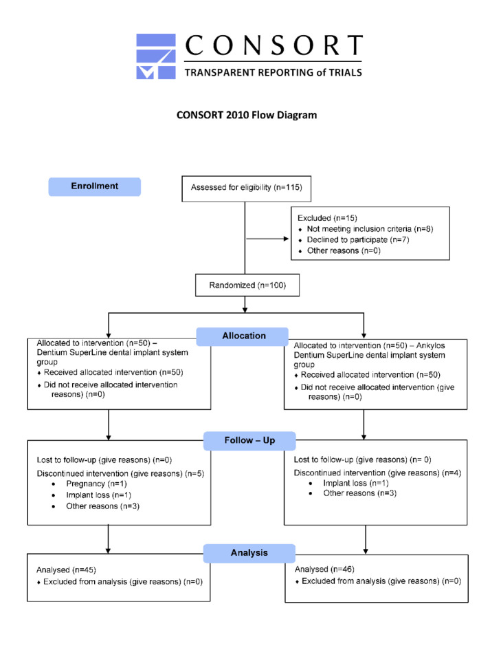
Material and methods: Gingival swabs were taken three times from 91 subjects. The first swab was taken before dental implant placement, the second swab 90 days after dental implant placement and the third swab 21 days following the healing abutment placement. DNA damage was analyzed using the micronucleus test. Tested dental implants with corresponding healing abutments were Ankylos and Dentium SuperLine.
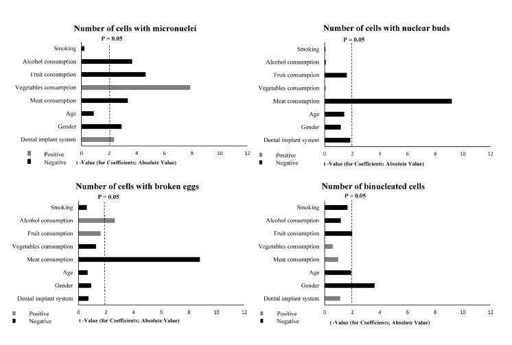
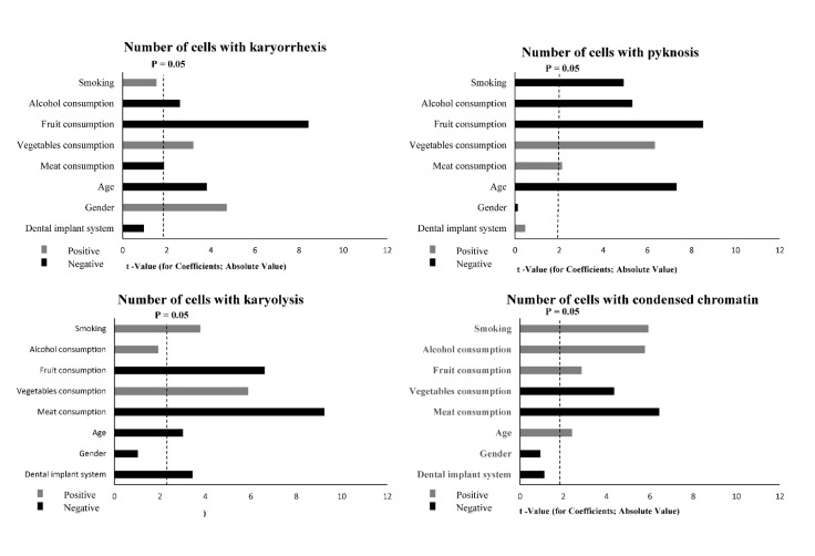
Results: Of all scored forms of cytogenetic damage in gingival cells of individuals after implementation of tested dental implant systems, only an increase in the number of binucleated cells (P ≤ 0.001) was significant in contrast to control values for both tested implant systems, 90 days after dental implant placement and 21 days following the healing abutment placement.
Conclusion: It may be concluded that there are no titanium-based implant dependent cytogenetic damage in gingival epithelial cells. A slight increase in cytogenetic damage has been observed but it is of no biological relevance and might be associated with healing abutment induced effect.
期刊介绍:
The Acta Stomatologica Croatica (ASCRO) is a leading scientific non-profit journal in the field of dental, oral and cranio-facial sciences during the past 44 years in Croatia. ASCRO publishes original scientific and clinical papers, preliminary communications, case reports, book reviews, letters to the editor and news. Review articles are published by invitation from the Editor-in-Chief by acclaimed professionals in distinct fields of dental medicine. All manuscripts are subjected to peer review process.


 分享
分享
 求助内容:
求助内容: 应助结果提醒方式:
 扫码关注我们
