{"title":"5-氟尿嘧啶诱导的幼鼠小脑外颗粒细胞损伤在时间过程中的变化。","authors":"Yuko Yamaguchi, Tsubasa Saito, Mizuho Takagi, Tomomi Nakazawa, Kazutoshi Tamura","doi":"10.1293/tox.2022-0003","DOIUrl":null,"url":null,"abstract":"<p><p>5-Fluorouracil (5-FU) is widely used as a chemotherapeutic agent that blocks DNA synthesis and replication by inhibiting thymidylate synthetase. This study aimed to elucidate 5-FU-induced changes in the external granular cells (EGCs) in the cerebellum of infant rats and the possible underlying mechanism. Six-day-old infant rats were injected subcutaneously with 40 mg/kg of 5-FU, and their cerebellums were examined at 6, 9, 12, and 24 h after treatment (HAT), and 2, 4, and 10 d after treatment (DAT). The width of the external granular layer (EGL) decreased from 24 HAT to 4 DAT in the 5-FU group compared to that in the control group. However, the width in the 5-FU group was comparable to that of the control group at 10 DAT. The number of apoptotic cells, cleaved caspase 3-labeling index (LI%), p21<sup>cip1</sup>-LI%, and expression levels of <i>p53</i>, <i>p21<sup>cip1</sup>,</i> and <i>Fas</i> mRNAs increased at 24 HAT. However, no changes were detected in the expression levels of <i>Puma</i> and <i>Bax</i> mRNAs at any time point. BrdU-LI% increased at 6 and 12 HAT but decreased at 24 HAT. The phospho-histone H3-LI% decreased from 6 HAT to 2 DAT. The width of the molecular layer decreased compared to that of the control group at 10 DAT. No differences were observed in Purkinje cell development. These results indicate that 5-FU inhibited cell proliferation by inducing apoptosis of EGCs via activation of Fas and caspase-3 without the involvement of the mitochondrial pathway and induced p53-dependent G1-S and G2-M phase arrest.</p>","PeriodicalId":17437,"journal":{"name":"Journal of Toxicologic Pathology","volume":"35 4","pages":"299-311"},"PeriodicalIF":0.9000,"publicationDate":"2022-10-01","publicationTypes":"Journal Article","fieldsOfStudy":null,"isOpenAccess":false,"openAccessPdf":"https://ftp.ncbi.nlm.nih.gov/pub/pmc/oa_pdf/51/f3/tox-35-299.PMC9647215.pdf","citationCount":"1","resultStr":"{\"title\":\"Changes in 5-Fluorouracil-induced external granular cell damage during the time-course of the developing cerebellum of infant rats.\",\"authors\":\"Yuko Yamaguchi, Tsubasa Saito, Mizuho Takagi, Tomomi Nakazawa, Kazutoshi Tamura\",\"doi\":\"10.1293/tox.2022-0003\",\"DOIUrl\":null,\"url\":null,\"abstract\":\"<p><p>5-Fluorouracil (5-FU) is widely used as a chemotherapeutic agent that blocks DNA synthesis and replication by inhibiting thymidylate synthetase. This study aimed to elucidate 5-FU-induced changes in the external granular cells (EGCs) in the cerebellum of infant rats and the possible underlying mechanism. Six-day-old infant rats were injected subcutaneously with 40 mg/kg of 5-FU, and their cerebellums were examined at 6, 9, 12, and 24 h after treatment (HAT), and 2, 4, and 10 d after treatment (DAT). The width of the external granular layer (EGL) decreased from 24 HAT to 4 DAT in the 5-FU group compared to that in the control group. However, the width in the 5-FU group was comparable to that of the control group at 10 DAT. The number of apoptotic cells, cleaved caspase 3-labeling index (LI%), p21<sup>cip1</sup>-LI%, and expression levels of <i>p53</i>, <i>p21<sup>cip1</sup>,</i> and <i>Fas</i> mRNAs increased at 24 HAT. However, no changes were detected in the expression levels of <i>Puma</i> and <i>Bax</i> mRNAs at any time point. BrdU-LI% increased at 6 and 12 HAT but decreased at 24 HAT. The phospho-histone H3-LI% decreased from 6 HAT to 2 DAT. The width of the molecular layer decreased compared to that of the control group at 10 DAT. No differences were observed in Purkinje cell development. These results indicate that 5-FU inhibited cell proliferation by inducing apoptosis of EGCs via activation of Fas and caspase-3 without the involvement of the mitochondrial pathway and induced p53-dependent G1-S and G2-M phase arrest.</p>\",\"PeriodicalId\":17437,\"journal\":{\"name\":\"Journal of Toxicologic Pathology\",\"volume\":\"35 4\",\"pages\":\"299-311\"},\"PeriodicalIF\":0.9000,\"publicationDate\":\"2022-10-01\",\"publicationTypes\":\"Journal Article\",\"fieldsOfStudy\":null,\"isOpenAccess\":false,\"openAccessPdf\":\"https://ftp.ncbi.nlm.nih.gov/pub/pmc/oa_pdf/51/f3/tox-35-299.PMC9647215.pdf\",\"citationCount\":\"1\",\"resultStr\":null,\"platform\":\"Semanticscholar\",\"paperid\":null,\"PeriodicalName\":\"Journal of Toxicologic Pathology\",\"FirstCategoryId\":\"3\",\"ListUrlMain\":\"https://doi.org/10.1293/tox.2022-0003\",\"RegionNum\":4,\"RegionCategory\":\"医学\",\"ArticlePicture\":[],\"TitleCN\":null,\"AbstractTextCN\":null,\"PMCID\":null,\"EPubDate\":\"2022/5/30 0:00:00\",\"PubModel\":\"Epub\",\"JCR\":\"Q4\",\"JCRName\":\"PATHOLOGY\",\"Score\":null,\"Total\":0}","platform":"Semanticscholar","paperid":null,"PeriodicalName":"Journal of Toxicologic Pathology","FirstCategoryId":"3","ListUrlMain":"https://doi.org/10.1293/tox.2022-0003","RegionNum":4,"RegionCategory":"医学","ArticlePicture":[],"TitleCN":null,"AbstractTextCN":null,"PMCID":null,"EPubDate":"2022/5/30 0:00:00","PubModel":"Epub","JCR":"Q4","JCRName":"PATHOLOGY","Score":null,"Total":0}
引用次数: 1
摘要
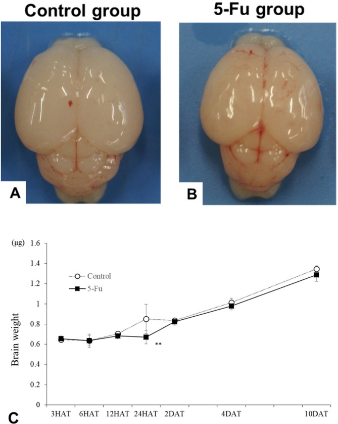
5-氟尿嘧啶(5-FU)是一种广泛使用的化疗药物,通过抑制胸苷酸合成酶来阻断DNA的合成和复制。本研究旨在阐明5- fu诱导的幼鼠小脑外颗粒细胞(EGCs)的变化及其可能的机制。6日龄幼鼠皮下注射5-FU 40 mg/kg,分别于给药后6、9、12、24 h (HAT)和给药后2、4、10 d (DAT)对其小脑进行检查。与对照组相比,5-FU组外颗粒层(EGL)宽度从24hat减小到4dat。然而,5-FU组在10dat时的宽度与对照组相当。凋亡细胞数量、裂解caspase 3标记指数(LI%)、p21cip1-LI%以及p53、p21cip1和Fas mrna的表达水平在24 HAT时增加。然而,在任何时间点Puma和Bax mrna的表达水平均未检测到变化。6和12 HAT时BrdU-LI%升高,24 HAT时降低。磷酸组蛋白H3-LI%由6 HAT降至2 DAT。与对照组相比,10 DAT时分子层宽度减小。浦肯野细胞发育无差异。这些结果表明,5-FU通过激活Fas和caspase-3而不参与线粒体途径诱导EGCs凋亡,并诱导p53依赖性G1-S和G2-M期阻滞,从而抑制细胞增殖。
Changes in 5-Fluorouracil-induced external granular cell damage during the time-course of the developing cerebellum of infant rats.
5-Fluorouracil (5-FU) is widely used as a chemotherapeutic agent that blocks DNA synthesis and replication by inhibiting thymidylate synthetase. This study aimed to elucidate 5-FU-induced changes in the external granular cells (EGCs) in the cerebellum of infant rats and the possible underlying mechanism. Six-day-old infant rats were injected subcutaneously with 40 mg/kg of 5-FU, and their cerebellums were examined at 6, 9, 12, and 24 h after treatment (HAT), and 2, 4, and 10 d after treatment (DAT). The width of the external granular layer (EGL) decreased from 24 HAT to 4 DAT in the 5-FU group compared to that in the control group. However, the width in the 5-FU group was comparable to that of the control group at 10 DAT. The number of apoptotic cells, cleaved caspase 3-labeling index (LI%), p21cip1-LI%, and expression levels of p53, p21cip1, and Fas mRNAs increased at 24 HAT. However, no changes were detected in the expression levels of Puma and Bax mRNAs at any time point. BrdU-LI% increased at 6 and 12 HAT but decreased at 24 HAT. The phospho-histone H3-LI% decreased from 6 HAT to 2 DAT. The width of the molecular layer decreased compared to that of the control group at 10 DAT. No differences were observed in Purkinje cell development. These results indicate that 5-FU inhibited cell proliferation by inducing apoptosis of EGCs via activation of Fas and caspase-3 without the involvement of the mitochondrial pathway and induced p53-dependent G1-S and G2-M phase arrest.
期刊介绍:
JTP is a scientific journal that publishes original studies in the field of toxicological pathology and in a wide variety of other related fields. The main scope of the journal is listed below.
Administrative Opinions of Policymakers and Regulatory Agencies
Adverse Events
Carcinogenesis
Data of A Predominantly Negative Nature
Drug-Induced Hematologic Toxicity
Embryological Pathology
High Throughput Pathology
Historical Data of Experimental Animals
Immunohistochemical Analysis
Molecular Pathology
Nomenclature of Lesions
Non-mammal Toxicity Study
Result or Lesion Induced by Chemicals of Which Names Hidden on Account of the Authors
Technology and Methodology Related to Toxicological Pathology
Tumor Pathology; Neoplasia and Hyperplasia
Ultrastructural Analysis
Use of Animal Models.


 分享
分享
 求助内容:
求助内容: 应助结果提醒方式:
 扫码关注我们
