Gabriel Conde , Mayumi Fernanda Aracati , Letícia Franchin Rodrigues , Susana Luporini de Oliveira , Camila Carlino da Costa , Ives Charlie-Silva , Thalles Fernando Rocha Ruiz , Sebastião Roberto Taboga , Marco Antonio de Andrade Belo
{"title":"罗非鱼疫苗递送系统中基于聚乳酸和维生素E的植入装置:生物相容性和生物降解研究","authors":"Gabriel Conde , Mayumi Fernanda Aracati , Letícia Franchin Rodrigues , Susana Luporini de Oliveira , Camila Carlino da Costa , Ives Charlie-Silva , Thalles Fernando Rocha Ruiz , Sebastião Roberto Taboga , Marco Antonio de Andrade Belo","doi":"10.1016/j.fsirep.2022.100060","DOIUrl":null,"url":null,"abstract":"<div><p>The use of Poly (lactic acid) (PLA) as a slow-release vehicle for vaccines has attracted the attention of researchers, since its insertion improves the uptake of them, and reduces side effects or by stimulating recruited defense cells, assisting immunity without the need for booster vaccine doses. Seeking to develop new strategies for the administration of drugs and vaccines in aquaculture, we evaluated the biocompatibility and biodegradation of polymeric PLA devices and PLA plus vitamin E devices, implanted through subcutaneous (SC) and intraperitoneal (IP) routes in Nile tilapia. To carry out this study, 84 male tilapia (initial 243.82 ± 56.74 g; final 400.71 ± 100.54 g) were randomly distributed in 3 tanks (<em>n</em> = 28 fish per treatment/tank). The devices were prepared in two formulations: neat PLA (containing 100% PLA) and PLAVE (PLA plus vitamin E) implanted using a commercial AnimalTag® applicator, and non-implanted fish (control). Fish were sampled 15, 30, 60, and 120 days post-implantation (DPI). Blood analysis was used to access blood cells and blood smear for differential leucocytes count. Serum biochemistry to evaluated changes in serum proteins and glycemia. Histopathological investigation using hematoxylin-eosin (H&E) was used to assess polymer-tissue interaction. Histochemistry and immunohistochemistry was used to detection immune cells and phagocytes in capsule, and analyses of melanomacrophage centers (MMCs) to morphometric evaluation and percentage amount of melanin, hemosiderin and lipofucsin pigments. Histopathological study revealed an increase of capsular formation and inflammatory cell infiltration in PLAVE-implanted tilapia through SC route (15 DPI). Tilapia implanted with PLAVE and PLA (SC) presented mast cells and eosinophilic granular cells during 15, 30, and 60 DPI, with a decrease in these cells in the fibrous capsule around the polymer at 120 DPI. PLAVE implanted tilapia SC at 60 DPI showed significantly phagocytosis points than other groups. Phagocytic cells (F4/80+) were observed near to biopolymers in phagocytosis sites. Lipofuscin at 120 DPI in spleen melanomacrophage centers were significantly high in PLAVE implanted tilapias when compared to fish with PLA implants and control. The serum biochemical study of tilapia did not reveal changes in cytotoxicity and liver function in implanted fish. The absence of side effects in hematological and biochemical findings, including the absence of mortality after device implantation, proves its clinical safety. PLA implants in tilapia have demonstrated biocompatibility, biodegradation, clinical safety, and excellent evolution of foreign body inflammatory responses.</p></div>","PeriodicalId":73029,"journal":{"name":"Fish and shellfish immunology reports","volume":"3 ","pages":"Article 100060"},"PeriodicalIF":2.8000,"publicationDate":"2022-12-01","publicationTypes":"Journal Article","fieldsOfStudy":null,"isOpenAccess":false,"openAccessPdf":"https://www.ncbi.nlm.nih.gov/pmc/articles/PMC9680062/pdf/","citationCount":"2","resultStr":"{\"title\":\"Device implant based on poly (lactic acid) with vitamin E for vaccine delivery system in Tilapia: Study for biocompatibility and biodegradation\",\"authors\":\"Gabriel Conde , Mayumi Fernanda Aracati , Letícia Franchin Rodrigues , Susana Luporini de Oliveira , Camila Carlino da Costa , Ives Charlie-Silva , Thalles Fernando Rocha Ruiz , Sebastião Roberto Taboga , Marco Antonio de Andrade Belo\",\"doi\":\"10.1016/j.fsirep.2022.100060\",\"DOIUrl\":null,\"url\":null,\"abstract\":\"<div><p>The use of Poly (lactic acid) (PLA) as a slow-release vehicle for vaccines has attracted the attention of researchers, since its insertion improves the uptake of them, and reduces side effects or by stimulating recruited defense cells, assisting immunity without the need for booster vaccine doses. Seeking to develop new strategies for the administration of drugs and vaccines in aquaculture, we evaluated the biocompatibility and biodegradation of polymeric PLA devices and PLA plus vitamin E devices, implanted through subcutaneous (SC) and intraperitoneal (IP) routes in Nile tilapia. To carry out this study, 84 male tilapia (initial 243.82 ± 56.74 g; final 400.71 ± 100.54 g) were randomly distributed in 3 tanks (<em>n</em> = 28 fish per treatment/tank). The devices were prepared in two formulations: neat PLA (containing 100% PLA) and PLAVE (PLA plus vitamin E) implanted using a commercial AnimalTag® applicator, and non-implanted fish (control). Fish were sampled 15, 30, 60, and 120 days post-implantation (DPI). Blood analysis was used to access blood cells and blood smear for differential leucocytes count. Serum biochemistry to evaluated changes in serum proteins and glycemia. Histopathological investigation using hematoxylin-eosin (H&E) was used to assess polymer-tissue interaction. Histochemistry and immunohistochemistry was used to detection immune cells and phagocytes in capsule, and analyses of melanomacrophage centers (MMCs) to morphometric evaluation and percentage amount of melanin, hemosiderin and lipofucsin pigments. Histopathological study revealed an increase of capsular formation and inflammatory cell infiltration in PLAVE-implanted tilapia through SC route (15 DPI). Tilapia implanted with PLAVE and PLA (SC) presented mast cells and eosinophilic granular cells during 15, 30, and 60 DPI, with a decrease in these cells in the fibrous capsule around the polymer at 120 DPI. PLAVE implanted tilapia SC at 60 DPI showed significantly phagocytosis points than other groups. Phagocytic cells (F4/80+) were observed near to biopolymers in phagocytosis sites. Lipofuscin at 120 DPI in spleen melanomacrophage centers were significantly high in PLAVE implanted tilapias when compared to fish with PLA implants and control. The serum biochemical study of tilapia did not reveal changes in cytotoxicity and liver function in implanted fish. The absence of side effects in hematological and biochemical findings, including the absence of mortality after device implantation, proves its clinical safety. PLA implants in tilapia have demonstrated biocompatibility, biodegradation, clinical safety, and excellent evolution of foreign body inflammatory responses.</p></div>\",\"PeriodicalId\":73029,\"journal\":{\"name\":\"Fish and shellfish immunology reports\",\"volume\":\"3 \",\"pages\":\"Article 100060\"},\"PeriodicalIF\":2.8000,\"publicationDate\":\"2022-12-01\",\"publicationTypes\":\"Journal Article\",\"fieldsOfStudy\":null,\"isOpenAccess\":false,\"openAccessPdf\":\"https://www.ncbi.nlm.nih.gov/pmc/articles/PMC9680062/pdf/\",\"citationCount\":\"2\",\"resultStr\":null,\"platform\":\"Semanticscholar\",\"paperid\":null,\"PeriodicalName\":\"Fish and shellfish immunology reports\",\"FirstCategoryId\":\"1085\",\"ListUrlMain\":\"https://www.sciencedirect.com/science/article/pii/S2667011922000111\",\"RegionNum\":0,\"RegionCategory\":null,\"ArticlePicture\":[],\"TitleCN\":null,\"AbstractTextCN\":null,\"PMCID\":null,\"EPubDate\":\"2022/7/3 0:00:00\",\"PubModel\":\"Epub\",\"JCR\":\"Q2\",\"JCRName\":\"FISHERIES\",\"Score\":null,\"Total\":0}","platform":"Semanticscholar","paperid":null,"PeriodicalName":"Fish and shellfish immunology reports","FirstCategoryId":"1085","ListUrlMain":"https://www.sciencedirect.com/science/article/pii/S2667011922000111","RegionNum":0,"RegionCategory":null,"ArticlePicture":[],"TitleCN":null,"AbstractTextCN":null,"PMCID":null,"EPubDate":"2022/7/3 0:00:00","PubModel":"Epub","JCR":"Q2","JCRName":"FISHERIES","Score":null,"Total":0}
Device implant based on poly (lactic acid) with vitamin E for vaccine delivery system in Tilapia: Study for biocompatibility and biodegradation
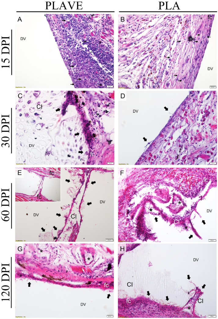
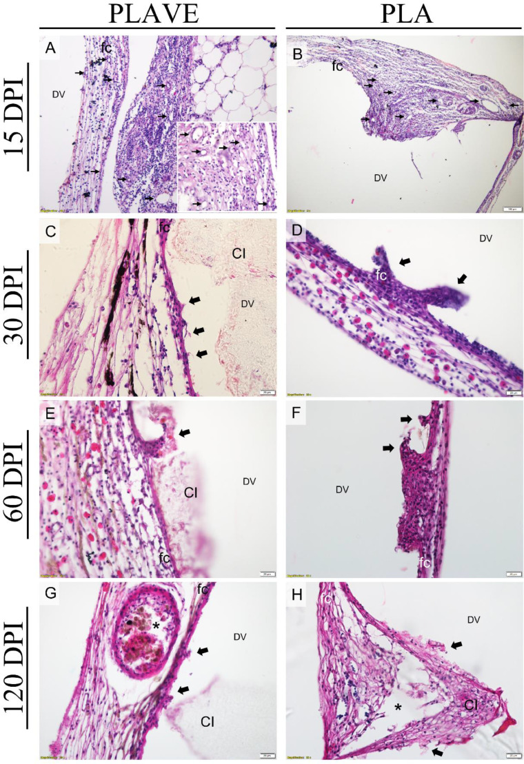
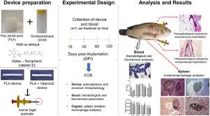
The use of Poly (lactic acid) (PLA) as a slow-release vehicle for vaccines has attracted the attention of researchers, since its insertion improves the uptake of them, and reduces side effects or by stimulating recruited defense cells, assisting immunity without the need for booster vaccine doses. Seeking to develop new strategies for the administration of drugs and vaccines in aquaculture, we evaluated the biocompatibility and biodegradation of polymeric PLA devices and PLA plus vitamin E devices, implanted through subcutaneous (SC) and intraperitoneal (IP) routes in Nile tilapia. To carry out this study, 84 male tilapia (initial 243.82 ± 56.74 g; final 400.71 ± 100.54 g) were randomly distributed in 3 tanks (n = 28 fish per treatment/tank). The devices were prepared in two formulations: neat PLA (containing 100% PLA) and PLAVE (PLA plus vitamin E) implanted using a commercial AnimalTag® applicator, and non-implanted fish (control). Fish were sampled 15, 30, 60, and 120 days post-implantation (DPI). Blood analysis was used to access blood cells and blood smear for differential leucocytes count. Serum biochemistry to evaluated changes in serum proteins and glycemia. Histopathological investigation using hematoxylin-eosin (H&E) was used to assess polymer-tissue interaction. Histochemistry and immunohistochemistry was used to detection immune cells and phagocytes in capsule, and analyses of melanomacrophage centers (MMCs) to morphometric evaluation and percentage amount of melanin, hemosiderin and lipofucsin pigments. Histopathological study revealed an increase of capsular formation and inflammatory cell infiltration in PLAVE-implanted tilapia through SC route (15 DPI). Tilapia implanted with PLAVE and PLA (SC) presented mast cells and eosinophilic granular cells during 15, 30, and 60 DPI, with a decrease in these cells in the fibrous capsule around the polymer at 120 DPI. PLAVE implanted tilapia SC at 60 DPI showed significantly phagocytosis points than other groups. Phagocytic cells (F4/80+) were observed near to biopolymers in phagocytosis sites. Lipofuscin at 120 DPI in spleen melanomacrophage centers were significantly high in PLAVE implanted tilapias when compared to fish with PLA implants and control. The serum biochemical study of tilapia did not reveal changes in cytotoxicity and liver function in implanted fish. The absence of side effects in hematological and biochemical findings, including the absence of mortality after device implantation, proves its clinical safety. PLA implants in tilapia have demonstrated biocompatibility, biodegradation, clinical safety, and excellent evolution of foreign body inflammatory responses.


 分享
分享

 求助内容:
求助内容: 应助结果提醒方式:
 扫码关注我们
