{"title":"Sprague-Dawley大鼠自发性睾丸网状腺瘤一例。","authors":"Masako Imaoka, Tetsuya Osawa, Kiyonori Kai, Yoshimi Tsuchiya","doi":"10.1293/tox.2022-0018","DOIUrl":null,"url":null,"abstract":"<p><p>A 104-week-old male CD (SD) rat exhibited enlargement of the left testis. Microscopically, this mass was demarcated from the testis by fibrous connective tissue and characterized by cystic dilatation with single-layered columnar cells and papillary proliferation connected to the solid growth area without clear boundaries. In the solid growth area, cells were dissected into irregular alveolar nests by scant fibrous tissue with small blood vessels. The nuclei of proliferating cells were variable in size and round- to oval-shaped, and their cytoplasm was pale or eosinophilic and sometimes contained vacuoles or eosinophilic granules. Immunohistochemically, the tumor cells were positive for vimentin and cytokeratin (CK) 7. Since CK7 was exclusively positive in the rete testis epithelium of the naïve rat, it was valuable to diagnose this tumor as rete testis-originated. Based on these results and the lack of apparent pleomorphism, mitotic figures, and metastasis, the present case was diagnosed as rete testis adenoma.</p>","PeriodicalId":17437,"journal":{"name":"Journal of Toxicologic Pathology","volume":"35 3","pages":"263-268"},"PeriodicalIF":0.9000,"publicationDate":"2022-07-01","publicationTypes":"Journal Article","fieldsOfStudy":null,"isOpenAccess":false,"openAccessPdf":"https://ftp.ncbi.nlm.nih.gov/pub/pmc/oa_pdf/bc/38/tox-35-263.PMC9255997.pdf","citationCount":"0","resultStr":"{\"title\":\"A case of spontaneous rete testis adenoma in a Sprague-Dawley rat.\",\"authors\":\"Masako Imaoka, Tetsuya Osawa, Kiyonori Kai, Yoshimi Tsuchiya\",\"doi\":\"10.1293/tox.2022-0018\",\"DOIUrl\":null,\"url\":null,\"abstract\":\"<p><p>A 104-week-old male CD (SD) rat exhibited enlargement of the left testis. Microscopically, this mass was demarcated from the testis by fibrous connective tissue and characterized by cystic dilatation with single-layered columnar cells and papillary proliferation connected to the solid growth area without clear boundaries. In the solid growth area, cells were dissected into irregular alveolar nests by scant fibrous tissue with small blood vessels. The nuclei of proliferating cells were variable in size and round- to oval-shaped, and their cytoplasm was pale or eosinophilic and sometimes contained vacuoles or eosinophilic granules. Immunohistochemically, the tumor cells were positive for vimentin and cytokeratin (CK) 7. Since CK7 was exclusively positive in the rete testis epithelium of the naïve rat, it was valuable to diagnose this tumor as rete testis-originated. Based on these results and the lack of apparent pleomorphism, mitotic figures, and metastasis, the present case was diagnosed as rete testis adenoma.</p>\",\"PeriodicalId\":17437,\"journal\":{\"name\":\"Journal of Toxicologic Pathology\",\"volume\":\"35 3\",\"pages\":\"263-268\"},\"PeriodicalIF\":0.9000,\"publicationDate\":\"2022-07-01\",\"publicationTypes\":\"Journal Article\",\"fieldsOfStudy\":null,\"isOpenAccess\":false,\"openAccessPdf\":\"https://ftp.ncbi.nlm.nih.gov/pub/pmc/oa_pdf/bc/38/tox-35-263.PMC9255997.pdf\",\"citationCount\":\"0\",\"resultStr\":null,\"platform\":\"Semanticscholar\",\"paperid\":null,\"PeriodicalName\":\"Journal of Toxicologic Pathology\",\"FirstCategoryId\":\"3\",\"ListUrlMain\":\"https://doi.org/10.1293/tox.2022-0018\",\"RegionNum\":4,\"RegionCategory\":\"医学\",\"ArticlePicture\":[],\"TitleCN\":null,\"AbstractTextCN\":null,\"PMCID\":null,\"EPubDate\":\"2022/5/7 0:00:00\",\"PubModel\":\"Epub\",\"JCR\":\"Q4\",\"JCRName\":\"PATHOLOGY\",\"Score\":null,\"Total\":0}","platform":"Semanticscholar","paperid":null,"PeriodicalName":"Journal of Toxicologic Pathology","FirstCategoryId":"3","ListUrlMain":"https://doi.org/10.1293/tox.2022-0018","RegionNum":4,"RegionCategory":"医学","ArticlePicture":[],"TitleCN":null,"AbstractTextCN":null,"PMCID":null,"EPubDate":"2022/5/7 0:00:00","PubModel":"Epub","JCR":"Q4","JCRName":"PATHOLOGY","Score":null,"Total":0}
A case of spontaneous rete testis adenoma in a Sprague-Dawley rat.
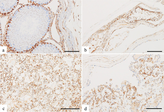
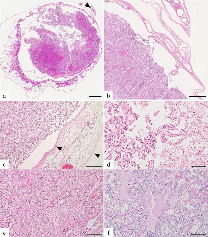
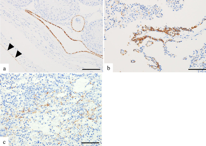
A 104-week-old male CD (SD) rat exhibited enlargement of the left testis. Microscopically, this mass was demarcated from the testis by fibrous connective tissue and characterized by cystic dilatation with single-layered columnar cells and papillary proliferation connected to the solid growth area without clear boundaries. In the solid growth area, cells were dissected into irregular alveolar nests by scant fibrous tissue with small blood vessels. The nuclei of proliferating cells were variable in size and round- to oval-shaped, and their cytoplasm was pale or eosinophilic and sometimes contained vacuoles or eosinophilic granules. Immunohistochemically, the tumor cells were positive for vimentin and cytokeratin (CK) 7. Since CK7 was exclusively positive in the rete testis epithelium of the naïve rat, it was valuable to diagnose this tumor as rete testis-originated. Based on these results and the lack of apparent pleomorphism, mitotic figures, and metastasis, the present case was diagnosed as rete testis adenoma.
期刊介绍:
JTP is a scientific journal that publishes original studies in the field of toxicological pathology and in a wide variety of other related fields. The main scope of the journal is listed below.
Administrative Opinions of Policymakers and Regulatory Agencies
Adverse Events
Carcinogenesis
Data of A Predominantly Negative Nature
Drug-Induced Hematologic Toxicity
Embryological Pathology
High Throughput Pathology
Historical Data of Experimental Animals
Immunohistochemical Analysis
Molecular Pathology
Nomenclature of Lesions
Non-mammal Toxicity Study
Result or Lesion Induced by Chemicals of Which Names Hidden on Account of the Authors
Technology and Methodology Related to Toxicological Pathology
Tumor Pathology; Neoplasia and Hyperplasia
Ultrastructural Analysis
Use of Animal Models.


 分享
分享
 求助内容:
求助内容: 应助结果提醒方式:
 扫码关注我们
