Chloé Chamard, Jerome J Maller, Nicolas Menjot, Eloi Debourdeau, Virginie Nael, Karen Ritchie, Isabelle Carriere, Vincent Daien
{"title":"在社区居住的老年人队列中视力与大脑皮质厚度的关系。","authors":"Chloé Chamard, Jerome J Maller, Nicolas Menjot, Eloi Debourdeau, Virginie Nael, Karen Ritchie, Isabelle Carriere, Vincent Daien","doi":"10.2147/EB.S358384","DOIUrl":null,"url":null,"abstract":"<p><strong>Purpose: </strong>Visual impairment is a major cause of disability and impairment of cognitive function in older people. Brain structural changes associated with visual function impairment are not well understood. The objective of this study was to assess the association between visual function and cortical thickness in older adults.</p><p><strong>Methods: </strong>Participants were selected from the French population-based ESPRIT cohort of 2259 community-dwelling adults ≥65 years old enrolled between 1999 and 2001. We considered visual function and brain MRI images at the 12-year follow-up in participants who were right-handed and free of dementia and/or stroke, randomly selected from the whole cohort. High-resolution structural T1-weighted brain scans acquired with a 3-Tesla scanner. Regional reconstruction and segmentation involved using the FreeSurfer image-analysis suite.</p><p><strong>Results: </strong>A total of 215 participants were included (mean [SD] age 81.8 [3.7] years; 53.0% women): 30 (14.0%) had central vision loss and 185 (86.0%) normal central vision. Vision loss was associated with thinner cortical thickness in the right insula (within the lateral sulcus of the brain) as compared with the control group (mean thickness 2.38 [0.04] vs 2.50 [0.03] mm, 4.8% thinning, p<sub>corrected</sub>= 0.04) after adjustment for age, sex, lifetime depression and cardiovascular disease.</p><p><strong>Conclusion: </strong>The present study describes a significant thinning of the right insular cortex in older adults with vision loss. The insula subserves a wide variety of functions in humans ranging from sensory and affective processing to high-level cognitive processing. Reduced insula thickness associated with vision loss may increase cognitive burden in the ageing brain.</p>","PeriodicalId":51844,"journal":{"name":"Eye and Brain","volume":" ","pages":"71-82"},"PeriodicalIF":2.4000,"publicationDate":"2022-07-14","publicationTypes":"Journal Article","fieldsOfStudy":null,"isOpenAccess":false,"openAccessPdf":"https://ftp.ncbi.nlm.nih.gov/pub/pmc/oa_pdf/fc/fd/eb-14-71.PMC9292457.pdf","citationCount":"0","resultStr":"{\"title\":\"Association Between Vision and Brain Cortical Thickness in a Community-Dwelling Elderly Cohort.\",\"authors\":\"Chloé Chamard, Jerome J Maller, Nicolas Menjot, Eloi Debourdeau, Virginie Nael, Karen Ritchie, Isabelle Carriere, Vincent Daien\",\"doi\":\"10.2147/EB.S358384\",\"DOIUrl\":null,\"url\":null,\"abstract\":\"<p><strong>Purpose: </strong>Visual impairment is a major cause of disability and impairment of cognitive function in older people. Brain structural changes associated with visual function impairment are not well understood. The objective of this study was to assess the association between visual function and cortical thickness in older adults.</p><p><strong>Methods: </strong>Participants were selected from the French population-based ESPRIT cohort of 2259 community-dwelling adults ≥65 years old enrolled between 1999 and 2001. We considered visual function and brain MRI images at the 12-year follow-up in participants who were right-handed and free of dementia and/or stroke, randomly selected from the whole cohort. High-resolution structural T1-weighted brain scans acquired with a 3-Tesla scanner. Regional reconstruction and segmentation involved using the FreeSurfer image-analysis suite.</p><p><strong>Results: </strong>A total of 215 participants were included (mean [SD] age 81.8 [3.7] years; 53.0% women): 30 (14.0%) had central vision loss and 185 (86.0%) normal central vision. Vision loss was associated with thinner cortical thickness in the right insula (within the lateral sulcus of the brain) as compared with the control group (mean thickness 2.38 [0.04] vs 2.50 [0.03] mm, 4.8% thinning, p<sub>corrected</sub>= 0.04) after adjustment for age, sex, lifetime depression and cardiovascular disease.</p><p><strong>Conclusion: </strong>The present study describes a significant thinning of the right insular cortex in older adults with vision loss. The insula subserves a wide variety of functions in humans ranging from sensory and affective processing to high-level cognitive processing. Reduced insula thickness associated with vision loss may increase cognitive burden in the ageing brain.</p>\",\"PeriodicalId\":51844,\"journal\":{\"name\":\"Eye and Brain\",\"volume\":\" \",\"pages\":\"71-82\"},\"PeriodicalIF\":2.4000,\"publicationDate\":\"2022-07-14\",\"publicationTypes\":\"Journal Article\",\"fieldsOfStudy\":null,\"isOpenAccess\":false,\"openAccessPdf\":\"https://ftp.ncbi.nlm.nih.gov/pub/pmc/oa_pdf/fc/fd/eb-14-71.PMC9292457.pdf\",\"citationCount\":\"0\",\"resultStr\":null,\"platform\":\"Semanticscholar\",\"paperid\":null,\"PeriodicalName\":\"Eye and Brain\",\"FirstCategoryId\":\"1085\",\"ListUrlMain\":\"https://doi.org/10.2147/EB.S358384\",\"RegionNum\":0,\"RegionCategory\":null,\"ArticlePicture\":[],\"TitleCN\":null,\"AbstractTextCN\":null,\"PMCID\":null,\"EPubDate\":\"2022/1/1 0:00:00\",\"PubModel\":\"eCollection\",\"JCR\":\"Q1\",\"JCRName\":\"OPHTHALMOLOGY\",\"Score\":null,\"Total\":0}","platform":"Semanticscholar","paperid":null,"PeriodicalName":"Eye and Brain","FirstCategoryId":"1085","ListUrlMain":"https://doi.org/10.2147/EB.S358384","RegionNum":0,"RegionCategory":null,"ArticlePicture":[],"TitleCN":null,"AbstractTextCN":null,"PMCID":null,"EPubDate":"2022/1/1 0:00:00","PubModel":"eCollection","JCR":"Q1","JCRName":"OPHTHALMOLOGY","Score":null,"Total":0}
Association Between Vision and Brain Cortical Thickness in a Community-Dwelling Elderly Cohort.
Purpose: Visual impairment is a major cause of disability and impairment of cognitive function in older people. Brain structural changes associated with visual function impairment are not well understood. The objective of this study was to assess the association between visual function and cortical thickness in older adults.
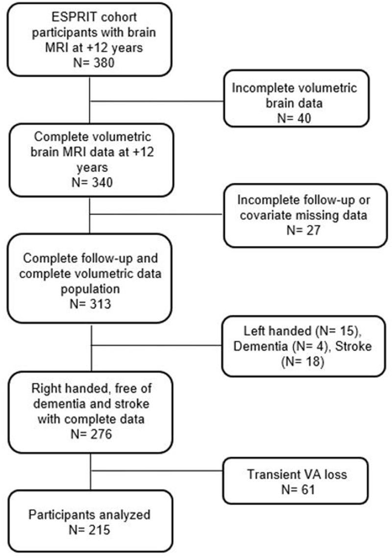
Methods: Participants were selected from the French population-based ESPRIT cohort of 2259 community-dwelling adults ≥65 years old enrolled between 1999 and 2001. We considered visual function and brain MRI images at the 12-year follow-up in participants who were right-handed and free of dementia and/or stroke, randomly selected from the whole cohort. High-resolution structural T1-weighted brain scans acquired with a 3-Tesla scanner. Regional reconstruction and segmentation involved using the FreeSurfer image-analysis suite.
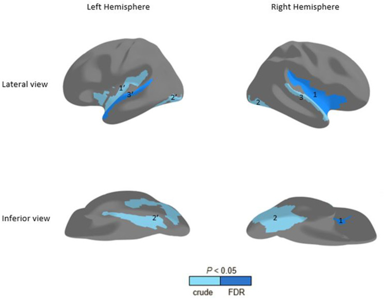
Results: A total of 215 participants were included (mean [SD] age 81.8 [3.7] years; 53.0% women): 30 (14.0%) had central vision loss and 185 (86.0%) normal central vision. Vision loss was associated with thinner cortical thickness in the right insula (within the lateral sulcus of the brain) as compared with the control group (mean thickness 2.38 [0.04] vs 2.50 [0.03] mm, 4.8% thinning, pcorrected= 0.04) after adjustment for age, sex, lifetime depression and cardiovascular disease.
Conclusion: The present study describes a significant thinning of the right insular cortex in older adults with vision loss. The insula subserves a wide variety of functions in humans ranging from sensory and affective processing to high-level cognitive processing. Reduced insula thickness associated with vision loss may increase cognitive burden in the ageing brain.
期刊介绍:
Eye and Brain is an international, peer-reviewed, open access journal focusing on basic research, clinical findings, and expert reviews in the field of visual science and neuro-ophthalmology. The journal’s unique focus is the link between two well-known visual centres, the eye and the brain, with an emphasis on the importance of such connections. All aspects of clinical and especially basic research on the visual system are addressed within the journal as well as significant future directions in vision research and therapeutic measures. This unique journal focuses on neurological aspects of vision – both physiological and pathological. The scope of the journal spans from the cornea to the associational visual cortex and all the visual centers in between. Topics range from basic biological mechanisms to therapeutic treatment, from simple organisms to humans, and utilizing techniques from molecular biology to behavior. The journal especially welcomes primary research articles or review papers that make the connection between the eye and the brain. Specific areas covered in the journal include: Physiology and pathophysiology of visual centers, Eye movement disorders and strabismus, Cellular, biochemical, and molecular features of the visual system, Structural and functional organization of the eye and of the visual cortex, Metabolic demands of the visual system, Diseases and disorders with neuro-ophthalmic manifestations, Clinical and experimental neuro-ophthalmology and visual system pathologies, Epidemiological studies.


 分享
分享
 求助内容:
求助内容: 应助结果提醒方式:
 扫码关注我们
