{"title":"蛋氨酸-正电子发射断层扫描模拟脑肿瘤的硬脑膜动静脉瘘1例。","authors":"Taketo Hanyu, Masahiro Nishihori, Takashi Izumi, Kazuya Motomura, Fumiharu Ohka, Shunsaku Goto, Yoshio Araki, Kinya Yokoyama, Kenji Uda, Ryuta Saito","doi":"10.2176/jns-nmc.2022-0055","DOIUrl":null,"url":null,"abstract":"<p><p>In this article, we report a case wherein a brain tumor was suspected based on computed tomography and magnetic resonance imaging findings. We made an initial diagnosis of malignant brain tumor based on methionine-positron emission tomography (PET) findings, but the correct diagnosis was dural arteriovenous fistula (DAVF). The patient was a 45-year-old man with DAVF who developed headache. Methionine-PET imaging showed high methionine uptake in the lesion. Although the tumor was strongly suspected from the findings of methionine-PET, the diagnosis of DAVF could be made correctly only by interpreting digital subtraction angiography and computed tomographic angiography. The findings of methionine-PET, which is considered useful in the diagnosis and denial of brain tumors, made the diagnosis of DAVF more difficult. The increased uptake of methionine-PET in DAVF is an important finding because, to our knowledge, this study is the first to report such finding. The results of this study might be useful for differential diagnoses when the diagnosis is uncertain.</p>","PeriodicalId":19260,"journal":{"name":"NMC Case Report Journal","volume":" ","pages":"289-294"},"PeriodicalIF":0.0000,"publicationDate":"2022-09-15","publicationTypes":"Journal Article","fieldsOfStudy":null,"isOpenAccess":false,"openAccessPdf":"https://ftp.ncbi.nlm.nih.gov/pub/pmc/oa_pdf/ac/80/2188-4226-9-0289.PMC9534565.pdf","citationCount":"1","resultStr":"{\"title\":\"Dural Arteriovenous Fistula Mimicking a Brain Tumor on Methionine-positron Emission Tomography: A Case Report.\",\"authors\":\"Taketo Hanyu, Masahiro Nishihori, Takashi Izumi, Kazuya Motomura, Fumiharu Ohka, Shunsaku Goto, Yoshio Araki, Kinya Yokoyama, Kenji Uda, Ryuta Saito\",\"doi\":\"10.2176/jns-nmc.2022-0055\",\"DOIUrl\":null,\"url\":null,\"abstract\":\"<p><p>In this article, we report a case wherein a brain tumor was suspected based on computed tomography and magnetic resonance imaging findings. We made an initial diagnosis of malignant brain tumor based on methionine-positron emission tomography (PET) findings, but the correct diagnosis was dural arteriovenous fistula (DAVF). The patient was a 45-year-old man with DAVF who developed headache. Methionine-PET imaging showed high methionine uptake in the lesion. Although the tumor was strongly suspected from the findings of methionine-PET, the diagnosis of DAVF could be made correctly only by interpreting digital subtraction angiography and computed tomographic angiography. The findings of methionine-PET, which is considered useful in the diagnosis and denial of brain tumors, made the diagnosis of DAVF more difficult. The increased uptake of methionine-PET in DAVF is an important finding because, to our knowledge, this study is the first to report such finding. The results of this study might be useful for differential diagnoses when the diagnosis is uncertain.</p>\",\"PeriodicalId\":19260,\"journal\":{\"name\":\"NMC Case Report Journal\",\"volume\":\" \",\"pages\":\"289-294\"},\"PeriodicalIF\":0.0000,\"publicationDate\":\"2022-09-15\",\"publicationTypes\":\"Journal Article\",\"fieldsOfStudy\":null,\"isOpenAccess\":false,\"openAccessPdf\":\"https://ftp.ncbi.nlm.nih.gov/pub/pmc/oa_pdf/ac/80/2188-4226-9-0289.PMC9534565.pdf\",\"citationCount\":\"1\",\"resultStr\":null,\"platform\":\"Semanticscholar\",\"paperid\":null,\"PeriodicalName\":\"NMC Case Report Journal\",\"FirstCategoryId\":\"1085\",\"ListUrlMain\":\"https://doi.org/10.2176/jns-nmc.2022-0055\",\"RegionNum\":0,\"RegionCategory\":null,\"ArticlePicture\":[],\"TitleCN\":null,\"AbstractTextCN\":null,\"PMCID\":null,\"EPubDate\":\"2022/1/1 0:00:00\",\"PubModel\":\"eCollection\",\"JCR\":\"\",\"JCRName\":\"\",\"Score\":null,\"Total\":0}","platform":"Semanticscholar","paperid":null,"PeriodicalName":"NMC Case Report Journal","FirstCategoryId":"1085","ListUrlMain":"https://doi.org/10.2176/jns-nmc.2022-0055","RegionNum":0,"RegionCategory":null,"ArticlePicture":[],"TitleCN":null,"AbstractTextCN":null,"PMCID":null,"EPubDate":"2022/1/1 0:00:00","PubModel":"eCollection","JCR":"","JCRName":"","Score":null,"Total":0}
Dural Arteriovenous Fistula Mimicking a Brain Tumor on Methionine-positron Emission Tomography: A Case Report.
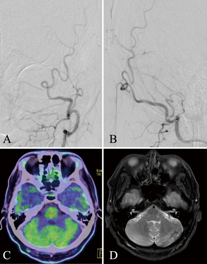
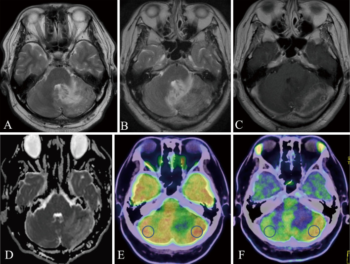
In this article, we report a case wherein a brain tumor was suspected based on computed tomography and magnetic resonance imaging findings. We made an initial diagnosis of malignant brain tumor based on methionine-positron emission tomography (PET) findings, but the correct diagnosis was dural arteriovenous fistula (DAVF). The patient was a 45-year-old man with DAVF who developed headache. Methionine-PET imaging showed high methionine uptake in the lesion. Although the tumor was strongly suspected from the findings of methionine-PET, the diagnosis of DAVF could be made correctly only by interpreting digital subtraction angiography and computed tomographic angiography. The findings of methionine-PET, which is considered useful in the diagnosis and denial of brain tumors, made the diagnosis of DAVF more difficult. The increased uptake of methionine-PET in DAVF is an important finding because, to our knowledge, this study is the first to report such finding. The results of this study might be useful for differential diagnoses when the diagnosis is uncertain.


 分享
分享

 求助内容:
求助内容: 应助结果提醒方式:
 扫码关注我们
