{"title":"子宫平滑肌肉瘤的形态学及辅助特征1例。","authors":"Mădălina Boșoteanu, Raluca Ioana Vodă, Mariana Așchie, Luana-Andreea Bosoteanu, Gabriela Izabela Bălțătescu","doi":"10.1177/2632010X221105224","DOIUrl":null,"url":null,"abstract":"<p><p>We report a rare case of giant uterine leiomyosarcoma in a postmenopausal woman, whose diagnosis was initially suspected at the evaluation of the abdominal efusion, and confirmed after the pathological examination of the uterus in association with the ancillary tests. The evaluation of the abdominal fluid showed single or clusters of malignant, round or spindle-shaped cells. On microscopic examination of the surgical specimen, a dense cell proliferation of spindle cells, with moderate to severe nuclear pleomorphism and significant mitotic activity was observed. Immunohistochemical evaluation demonstrated the loss of myocytic differentiation by focal, weakly positive expression of smooth muscle actin and desmin. The data presented in this case emphasize the relevance of the cytological examination, although the latter has only indicative value, especially since it is an aggressive tumor, frequently associated with mutant expression of p53. In our case, the first indication of the presence of uterine sarcoma was given by the presence of atypical cells in the peritoneal fluid.</p>","PeriodicalId":53204,"journal":{"name":"Clinical Pathology","volume":" ","pages":"2632010X221105224"},"PeriodicalIF":1.9000,"publicationDate":"2022-06-27","publicationTypes":"Journal Article","fieldsOfStudy":null,"isOpenAccess":false,"openAccessPdf":"https://ftp.ncbi.nlm.nih.gov/pub/pmc/oa_pdf/7b/fb/10.1177_2632010X221105224.PMC9240338.pdf","citationCount":"1","resultStr":"{\"title\":\"Morphological and Ancillary Features of Uterine Leiomyosarcoma: Case Report.\",\"authors\":\"Mădălina Boșoteanu, Raluca Ioana Vodă, Mariana Așchie, Luana-Andreea Bosoteanu, Gabriela Izabela Bălțătescu\",\"doi\":\"10.1177/2632010X221105224\",\"DOIUrl\":null,\"url\":null,\"abstract\":\"<p><p>We report a rare case of giant uterine leiomyosarcoma in a postmenopausal woman, whose diagnosis was initially suspected at the evaluation of the abdominal efusion, and confirmed after the pathological examination of the uterus in association with the ancillary tests. The evaluation of the abdominal fluid showed single or clusters of malignant, round or spindle-shaped cells. On microscopic examination of the surgical specimen, a dense cell proliferation of spindle cells, with moderate to severe nuclear pleomorphism and significant mitotic activity was observed. Immunohistochemical evaluation demonstrated the loss of myocytic differentiation by focal, weakly positive expression of smooth muscle actin and desmin. The data presented in this case emphasize the relevance of the cytological examination, although the latter has only indicative value, especially since it is an aggressive tumor, frequently associated with mutant expression of p53. In our case, the first indication of the presence of uterine sarcoma was given by the presence of atypical cells in the peritoneal fluid.</p>\",\"PeriodicalId\":53204,\"journal\":{\"name\":\"Clinical Pathology\",\"volume\":\" \",\"pages\":\"2632010X221105224\"},\"PeriodicalIF\":1.9000,\"publicationDate\":\"2022-06-27\",\"publicationTypes\":\"Journal Article\",\"fieldsOfStudy\":null,\"isOpenAccess\":false,\"openAccessPdf\":\"https://ftp.ncbi.nlm.nih.gov/pub/pmc/oa_pdf/7b/fb/10.1177_2632010X221105224.PMC9240338.pdf\",\"citationCount\":\"1\",\"resultStr\":null,\"platform\":\"Semanticscholar\",\"paperid\":null,\"PeriodicalName\":\"Clinical Pathology\",\"FirstCategoryId\":\"1085\",\"ListUrlMain\":\"https://doi.org/10.1177/2632010X221105224\",\"RegionNum\":0,\"RegionCategory\":null,\"ArticlePicture\":[],\"TitleCN\":null,\"AbstractTextCN\":null,\"PMCID\":null,\"EPubDate\":\"2022/1/1 0:00:00\",\"PubModel\":\"eCollection\",\"JCR\":\"Q3\",\"JCRName\":\"PATHOLOGY\",\"Score\":null,\"Total\":0}","platform":"Semanticscholar","paperid":null,"PeriodicalName":"Clinical Pathology","FirstCategoryId":"1085","ListUrlMain":"https://doi.org/10.1177/2632010X221105224","RegionNum":0,"RegionCategory":null,"ArticlePicture":[],"TitleCN":null,"AbstractTextCN":null,"PMCID":null,"EPubDate":"2022/1/1 0:00:00","PubModel":"eCollection","JCR":"Q3","JCRName":"PATHOLOGY","Score":null,"Total":0}
Morphological and Ancillary Features of Uterine Leiomyosarcoma: Case Report.
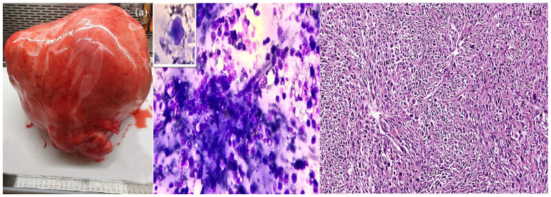
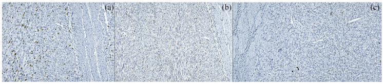
We report a rare case of giant uterine leiomyosarcoma in a postmenopausal woman, whose diagnosis was initially suspected at the evaluation of the abdominal efusion, and confirmed after the pathological examination of the uterus in association with the ancillary tests. The evaluation of the abdominal fluid showed single or clusters of malignant, round or spindle-shaped cells. On microscopic examination of the surgical specimen, a dense cell proliferation of spindle cells, with moderate to severe nuclear pleomorphism and significant mitotic activity was observed. Immunohistochemical evaluation demonstrated the loss of myocytic differentiation by focal, weakly positive expression of smooth muscle actin and desmin. The data presented in this case emphasize the relevance of the cytological examination, although the latter has only indicative value, especially since it is an aggressive tumor, frequently associated with mutant expression of p53. In our case, the first indication of the presence of uterine sarcoma was given by the presence of atypical cells in the peritoneal fluid.


 分享
分享
 求助内容:
求助内容: 应助结果提醒方式:
 扫码关注我们
