Xiaohui Dai, Jiao Chen, Hanmin Liu, Lin Wu, Fumin Zhao
{"title":"降落伞二尖瓣的产前诊断能否实现?胎儿降落伞二尖瓣1例报告。","authors":"Xiaohui Dai, Jiao Chen, Hanmin Liu, Lin Wu, Fumin Zhao","doi":"10.1186/s12947-022-00288-z","DOIUrl":null,"url":null,"abstract":"<p><p>Parachute mitral valve (PMV) is a common form of congenital mitral stenosis and is difficult to diagnose prenatally. This report describes a fetal case of PMV with coarctation of the aorta that was diagnosed at 25 weeks' gestation by echocardiography and confirmed at autopsy. We describe the ultrasonographic features in this case and present a useful sign for making a prenatal diagnosis of PMV.</p>","PeriodicalId":9613,"journal":{"name":"Cardiovascular Ultrasound","volume":" ","pages":"16"},"PeriodicalIF":1.6000,"publicationDate":"2022-07-08","publicationTypes":"Journal Article","fieldsOfStudy":null,"isOpenAccess":false,"openAccessPdf":"https://www.ncbi.nlm.nih.gov/pmc/articles/PMC9264502/pdf/","citationCount":"0","resultStr":"{\"title\":\"Can prenatal diagnosis of parachute mitral valve be achieved? A case report of fetal parachute mitral valve.\",\"authors\":\"Xiaohui Dai, Jiao Chen, Hanmin Liu, Lin Wu, Fumin Zhao\",\"doi\":\"10.1186/s12947-022-00288-z\",\"DOIUrl\":null,\"url\":null,\"abstract\":\"<p><p>Parachute mitral valve (PMV) is a common form of congenital mitral stenosis and is difficult to diagnose prenatally. This report describes a fetal case of PMV with coarctation of the aorta that was diagnosed at 25 weeks' gestation by echocardiography and confirmed at autopsy. We describe the ultrasonographic features in this case and present a useful sign for making a prenatal diagnosis of PMV.</p>\",\"PeriodicalId\":9613,\"journal\":{\"name\":\"Cardiovascular Ultrasound\",\"volume\":\" \",\"pages\":\"16\"},\"PeriodicalIF\":1.6000,\"publicationDate\":\"2022-07-08\",\"publicationTypes\":\"Journal Article\",\"fieldsOfStudy\":null,\"isOpenAccess\":false,\"openAccessPdf\":\"https://www.ncbi.nlm.nih.gov/pmc/articles/PMC9264502/pdf/\",\"citationCount\":\"0\",\"resultStr\":null,\"platform\":\"Semanticscholar\",\"paperid\":null,\"PeriodicalName\":\"Cardiovascular Ultrasound\",\"FirstCategoryId\":\"3\",\"ListUrlMain\":\"https://doi.org/10.1186/s12947-022-00288-z\",\"RegionNum\":3,\"RegionCategory\":\"医学\",\"ArticlePicture\":[],\"TitleCN\":null,\"AbstractTextCN\":null,\"PMCID\":null,\"EPubDate\":\"\",\"PubModel\":\"\",\"JCR\":\"Q3\",\"JCRName\":\"CARDIAC & CARDIOVASCULAR SYSTEMS\",\"Score\":null,\"Total\":0}","platform":"Semanticscholar","paperid":null,"PeriodicalName":"Cardiovascular Ultrasound","FirstCategoryId":"3","ListUrlMain":"https://doi.org/10.1186/s12947-022-00288-z","RegionNum":3,"RegionCategory":"医学","ArticlePicture":[],"TitleCN":null,"AbstractTextCN":null,"PMCID":null,"EPubDate":"","PubModel":"","JCR":"Q3","JCRName":"CARDIAC & CARDIOVASCULAR SYSTEMS","Score":null,"Total":0}
Can prenatal diagnosis of parachute mitral valve be achieved? A case report of fetal parachute mitral valve.
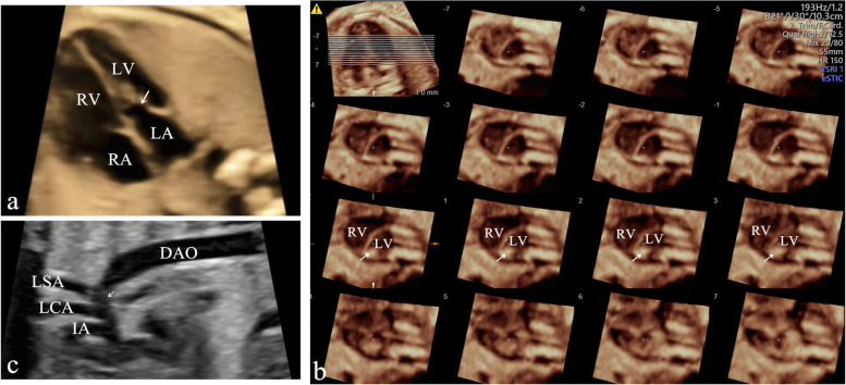
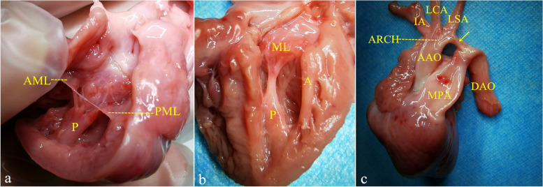
Parachute mitral valve (PMV) is a common form of congenital mitral stenosis and is difficult to diagnose prenatally. This report describes a fetal case of PMV with coarctation of the aorta that was diagnosed at 25 weeks' gestation by echocardiography and confirmed at autopsy. We describe the ultrasonographic features in this case and present a useful sign for making a prenatal diagnosis of PMV.
期刊介绍:
Cardiovascular Ultrasound is an online journal, publishing peer-reviewed: original research; authoritative reviews; case reports on challenging and/or unusual diagnostic aspects; and expert opinions on new techniques and technologies. We are particularly interested in articles that include relevant images or video files, which provide an additional dimension to published articles and enhance understanding.
As an open access journal, Cardiovascular Ultrasound ensures high visibility for authors in addition to providing an up-to-date and freely available resource for the community. The journal welcomes discussion, and provides a forum for publishing opinion and debate ranging from biology to engineering to clinical echocardiography, with both speed and versatility.


 分享
分享
 求助内容:
求助内容: 应助结果提醒方式:
 扫码关注我们
