Albert John Bromeo, Sweet Jorlene Lerit, Patricia Grulla-Quilendrino, George Michael Sosuan, Edgar Leuenberger
{"title":"小梁切除术后视网膜中央动静脉联合闭塞。","authors":"Albert John Bromeo, Sweet Jorlene Lerit, Patricia Grulla-Quilendrino, George Michael Sosuan, Edgar Leuenberger","doi":"10.3205/oc000205","DOIUrl":null,"url":null,"abstract":"<p><p>Retinal vascular events may occur as rare complications of glaucoma procedures due to various factors, including exacerbation of ischemia in patients with pre-existing vascular comorbidities, toxic effect of mitomycin-C, and decompression retinopathy. We present the case of a 47-year-old hypertensive male who underwent trabeculectomy for advanced glaucoma in his right eye. At 3 weeks postoperatively, he presented with a drop in visual acuity to light perception with a spike in intraocular pressure. On examination, there was increased bleb vascularity as well as rubeosis. Fundoscopy revealed findings consistent with both central retinal artery occlusion and central retinal vein occlusion. Combined central retinal artery and vein occlusion is a rare retinal vascular condition. Neovascular glaucoma can occur as a sequelae of the ischemic process in the retina. Despite treatment, there is a poor visual prognosis, with the affected eye usually becoming blind from optic atrophy and neovascularization.</p>","PeriodicalId":73178,"journal":{"name":"GMS ophthalmology cases","volume":" ","pages":"Doc18"},"PeriodicalIF":0.0000,"publicationDate":"2022-06-28","publicationTypes":"Journal Article","fieldsOfStudy":null,"isOpenAccess":false,"openAccessPdf":"https://www.ncbi.nlm.nih.gov/pmc/articles/PMC9285110/pdf/","citationCount":"1","resultStr":"{\"title\":\"Combined central retinal artery and vein occlusion following trabeculectomy.\",\"authors\":\"Albert John Bromeo, Sweet Jorlene Lerit, Patricia Grulla-Quilendrino, George Michael Sosuan, Edgar Leuenberger\",\"doi\":\"10.3205/oc000205\",\"DOIUrl\":null,\"url\":null,\"abstract\":\"<p><p>Retinal vascular events may occur as rare complications of glaucoma procedures due to various factors, including exacerbation of ischemia in patients with pre-existing vascular comorbidities, toxic effect of mitomycin-C, and decompression retinopathy. We present the case of a 47-year-old hypertensive male who underwent trabeculectomy for advanced glaucoma in his right eye. At 3 weeks postoperatively, he presented with a drop in visual acuity to light perception with a spike in intraocular pressure. On examination, there was increased bleb vascularity as well as rubeosis. Fundoscopy revealed findings consistent with both central retinal artery occlusion and central retinal vein occlusion. Combined central retinal artery and vein occlusion is a rare retinal vascular condition. Neovascular glaucoma can occur as a sequelae of the ischemic process in the retina. Despite treatment, there is a poor visual prognosis, with the affected eye usually becoming blind from optic atrophy and neovascularization.</p>\",\"PeriodicalId\":73178,\"journal\":{\"name\":\"GMS ophthalmology cases\",\"volume\":\" \",\"pages\":\"Doc18\"},\"PeriodicalIF\":0.0000,\"publicationDate\":\"2022-06-28\",\"publicationTypes\":\"Journal Article\",\"fieldsOfStudy\":null,\"isOpenAccess\":false,\"openAccessPdf\":\"https://www.ncbi.nlm.nih.gov/pmc/articles/PMC9285110/pdf/\",\"citationCount\":\"1\",\"resultStr\":null,\"platform\":\"Semanticscholar\",\"paperid\":null,\"PeriodicalName\":\"GMS ophthalmology cases\",\"FirstCategoryId\":\"1085\",\"ListUrlMain\":\"https://doi.org/10.3205/oc000205\",\"RegionNum\":0,\"RegionCategory\":null,\"ArticlePicture\":[],\"TitleCN\":null,\"AbstractTextCN\":null,\"PMCID\":null,\"EPubDate\":\"2022/1/1 0:00:00\",\"PubModel\":\"eCollection\",\"JCR\":\"\",\"JCRName\":\"\",\"Score\":null,\"Total\":0}","platform":"Semanticscholar","paperid":null,"PeriodicalName":"GMS ophthalmology cases","FirstCategoryId":"1085","ListUrlMain":"https://doi.org/10.3205/oc000205","RegionNum":0,"RegionCategory":null,"ArticlePicture":[],"TitleCN":null,"AbstractTextCN":null,"PMCID":null,"EPubDate":"2022/1/1 0:00:00","PubModel":"eCollection","JCR":"","JCRName":"","Score":null,"Total":0}
Combined central retinal artery and vein occlusion following trabeculectomy.
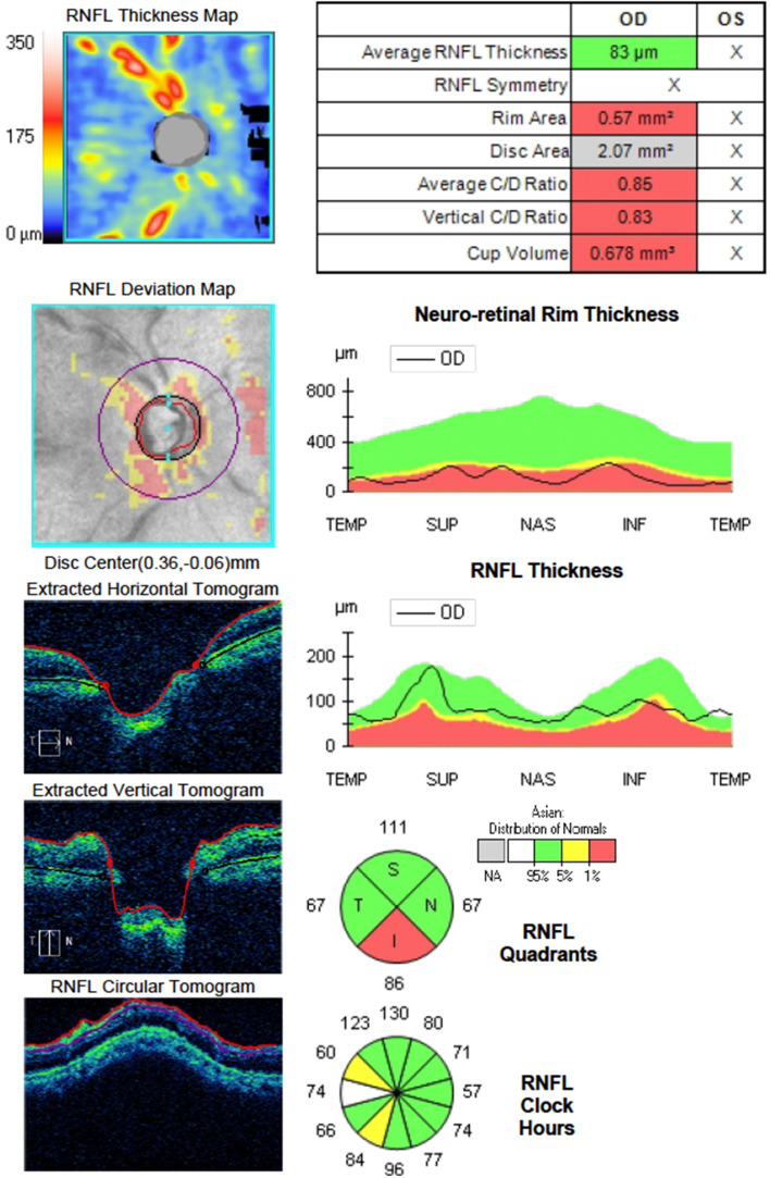
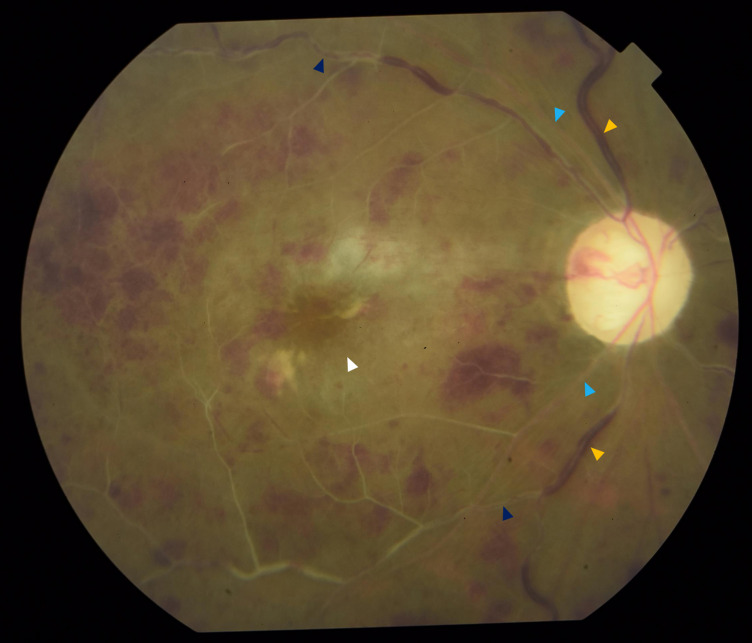
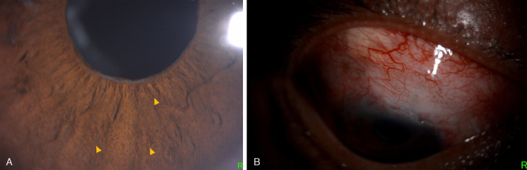
Retinal vascular events may occur as rare complications of glaucoma procedures due to various factors, including exacerbation of ischemia in patients with pre-existing vascular comorbidities, toxic effect of mitomycin-C, and decompression retinopathy. We present the case of a 47-year-old hypertensive male who underwent trabeculectomy for advanced glaucoma in his right eye. At 3 weeks postoperatively, he presented with a drop in visual acuity to light perception with a spike in intraocular pressure. On examination, there was increased bleb vascularity as well as rubeosis. Fundoscopy revealed findings consistent with both central retinal artery occlusion and central retinal vein occlusion. Combined central retinal artery and vein occlusion is a rare retinal vascular condition. Neovascular glaucoma can occur as a sequelae of the ischemic process in the retina. Despite treatment, there is a poor visual prognosis, with the affected eye usually becoming blind from optic atrophy and neovascularization.


 分享
分享

 求助内容:
求助内容: 应助结果提醒方式:
 扫码关注我们
