Kamila Wojtowicz, Tomasz Góra, Paweł Guzik, Magdalena Harpula, Paweł Chechliński, Ewelina Wolak, Aleksandra Stryjkowska-Góra
{"title":"子宫肌瘤和肉瘤-临床和超声特征和鉴别诊断使用脉冲和彩色多普勒技术。","authors":"Kamila Wojtowicz, Tomasz Góra, Paweł Guzik, Magdalena Harpula, Paweł Chechliński, Ewelina Wolak, Aleksandra Stryjkowska-Góra","doi":"10.15557/JoU.2022.0017","DOIUrl":null,"url":null,"abstract":"<p><p>Uterine tumors are a challenge encountered by every gynecologist in clinical practice. In the era of increasing incidence of endometrial cancer in the general population of women at reproductive age, compared to other genital malignancies, we should not forget about other tumors originating from the mucous and muscular layer of the uterus. Clear ultrasonographic differentiation of uterine tumors into benign (myomas) and malignant (sarcomas) lesions may sometimes prove impossible. Myomas, the most common uterine tumors, are characterized by discrete vascularization on color Doppler and high blood flow velocity as well as the lack of early diastolic notch on Doppler ultrasound. Sarcomas, on the other hand, show characteristic rich vascularization. Rapid tumor growth should also be noted when making the diagnosis. There are multiple known causes of uterine tumors. So far, no clear Doppler flow markers have been identified to characterize benign and malignant lesions.</p>","PeriodicalId":45612,"journal":{"name":"Journal of Ultrasonography","volume":"22 89","pages":"100-108"},"PeriodicalIF":1.5000,"publicationDate":"2022-04-27","publicationTypes":"Journal Article","fieldsOfStudy":null,"isOpenAccess":false,"openAccessPdf":"https://ftp.ncbi.nlm.nih.gov/pub/pmc/oa_pdf/b8/5c/jou-22-100.PMC9231509.pdf","citationCount":"4","resultStr":"{\"title\":\"Uterine Myomas and Sarcomas - Clinical and Ultrasound Characteristics and Differential Diagnosis Using Pulsed and Color Doppler Techniques.\",\"authors\":\"Kamila Wojtowicz, Tomasz Góra, Paweł Guzik, Magdalena Harpula, Paweł Chechliński, Ewelina Wolak, Aleksandra Stryjkowska-Góra\",\"doi\":\"10.15557/JoU.2022.0017\",\"DOIUrl\":null,\"url\":null,\"abstract\":\"<p><p>Uterine tumors are a challenge encountered by every gynecologist in clinical practice. In the era of increasing incidence of endometrial cancer in the general population of women at reproductive age, compared to other genital malignancies, we should not forget about other tumors originating from the mucous and muscular layer of the uterus. Clear ultrasonographic differentiation of uterine tumors into benign (myomas) and malignant (sarcomas) lesions may sometimes prove impossible. Myomas, the most common uterine tumors, are characterized by discrete vascularization on color Doppler and high blood flow velocity as well as the lack of early diastolic notch on Doppler ultrasound. Sarcomas, on the other hand, show characteristic rich vascularization. Rapid tumor growth should also be noted when making the diagnosis. There are multiple known causes of uterine tumors. So far, no clear Doppler flow markers have been identified to characterize benign and malignant lesions.</p>\",\"PeriodicalId\":45612,\"journal\":{\"name\":\"Journal of Ultrasonography\",\"volume\":\"22 89\",\"pages\":\"100-108\"},\"PeriodicalIF\":1.5000,\"publicationDate\":\"2022-04-27\",\"publicationTypes\":\"Journal Article\",\"fieldsOfStudy\":null,\"isOpenAccess\":false,\"openAccessPdf\":\"https://ftp.ncbi.nlm.nih.gov/pub/pmc/oa_pdf/b8/5c/jou-22-100.PMC9231509.pdf\",\"citationCount\":\"4\",\"resultStr\":null,\"platform\":\"Semanticscholar\",\"paperid\":null,\"PeriodicalName\":\"Journal of Ultrasonography\",\"FirstCategoryId\":\"1085\",\"ListUrlMain\":\"https://doi.org/10.15557/JoU.2022.0017\",\"RegionNum\":0,\"RegionCategory\":null,\"ArticlePicture\":[],\"TitleCN\":null,\"AbstractTextCN\":null,\"PMCID\":null,\"EPubDate\":\"2022/4/1 0:00:00\",\"PubModel\":\"eCollection\",\"JCR\":\"Q3\",\"JCRName\":\"RADIOLOGY, NUCLEAR MEDICINE & MEDICAL IMAGING\",\"Score\":null,\"Total\":0}","platform":"Semanticscholar","paperid":null,"PeriodicalName":"Journal of Ultrasonography","FirstCategoryId":"1085","ListUrlMain":"https://doi.org/10.15557/JoU.2022.0017","RegionNum":0,"RegionCategory":null,"ArticlePicture":[],"TitleCN":null,"AbstractTextCN":null,"PMCID":null,"EPubDate":"2022/4/1 0:00:00","PubModel":"eCollection","JCR":"Q3","JCRName":"RADIOLOGY, NUCLEAR MEDICINE & MEDICAL IMAGING","Score":null,"Total":0}
Uterine Myomas and Sarcomas - Clinical and Ultrasound Characteristics and Differential Diagnosis Using Pulsed and Color Doppler Techniques.
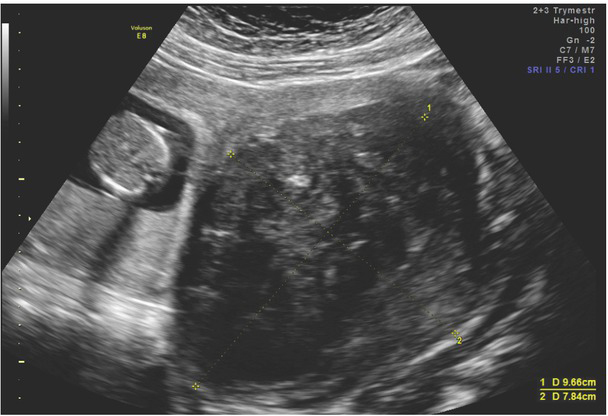
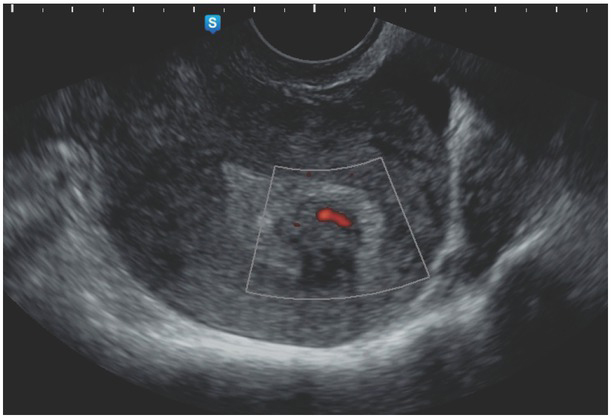
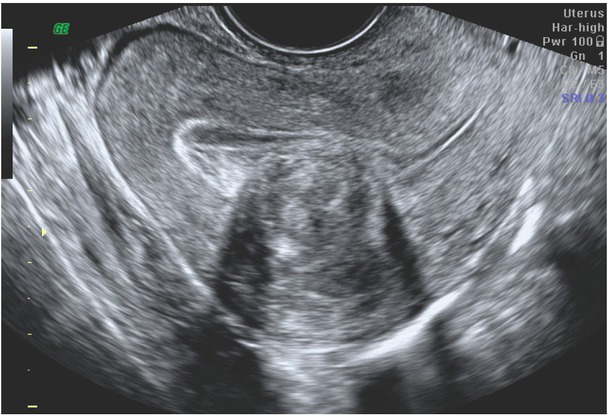
Uterine tumors are a challenge encountered by every gynecologist in clinical practice. In the era of increasing incidence of endometrial cancer in the general population of women at reproductive age, compared to other genital malignancies, we should not forget about other tumors originating from the mucous and muscular layer of the uterus. Clear ultrasonographic differentiation of uterine tumors into benign (myomas) and malignant (sarcomas) lesions may sometimes prove impossible. Myomas, the most common uterine tumors, are characterized by discrete vascularization on color Doppler and high blood flow velocity as well as the lack of early diastolic notch on Doppler ultrasound. Sarcomas, on the other hand, show characteristic rich vascularization. Rapid tumor growth should also be noted when making the diagnosis. There are multiple known causes of uterine tumors. So far, no clear Doppler flow markers have been identified to characterize benign and malignant lesions.


 分享
分享
 求助内容:
求助内容: 应助结果提醒方式:
 扫码关注我们
