{"title":"斑马鱼Slit2和Slit3共同作用调节视网膜轴突中线交叉。","authors":"Camila Davison, Gabriela Bedó, Flavio R Zolessi","doi":"10.3390/jdb10040041","DOIUrl":null,"url":null,"abstract":"<p><p>Slit-Robo signaling regulates midline crossing of commissural axons in different systems. In zebrafish, all retinofugal axons cross at the optic chiasm to innervate the contralateral tectum. Here, the mutant for the Robo2 receptor presents severe axon guidance defects, which were not completely reproduced in a Slit2 ligand null mutant. Since <i>slit3</i> is also expressed around this area at the stage of axon crossing, we decided to analyze the possibility that it collaborates with Slit2 in this process. We found that the disruption of <i>slit3</i> expression by sgRNA-Cas9 injection caused similar, albeit slightly milder, defects than those of the <i>slit2</i> mutant, while the same treatment in the <i>slit2-/-<sup>mz</sup></i> background caused much more severe defects, comparable to those observed in <i>robo2</i> mutants. Tracking analysis of in vivo time-lapse experiments indicated differential but complementary functions of these secreted factors in the correction of axon turn errors around the optic chiasm. Interestingly, RT-qPCR analysis showed a mild increase in <i>slit2</i> expression in <i>slit3</i>-deficient embryos, but not the opposite. Our observations support the previously proposed \"repulsive channel\" model for Slit-Robo action at the optic chiasm, with both Slits acting in different manners, most probably relating to their different spatial expression patterns.</p>","PeriodicalId":15563,"journal":{"name":"Journal of Developmental Biology","volume":" ","pages":""},"PeriodicalIF":2.5000,"publicationDate":"2022-09-23","publicationTypes":"Journal Article","fieldsOfStudy":null,"isOpenAccess":false,"openAccessPdf":"https://www.ncbi.nlm.nih.gov/pmc/articles/PMC9590056/pdf/","citationCount":"0","resultStr":"{\"title\":\"Zebrafish Slit2 and Slit3 Act Together to Regulate Retinal Axon Crossing at the Midline.\",\"authors\":\"Camila Davison, Gabriela Bedó, Flavio R Zolessi\",\"doi\":\"10.3390/jdb10040041\",\"DOIUrl\":null,\"url\":null,\"abstract\":\"<p><p>Slit-Robo signaling regulates midline crossing of commissural axons in different systems. In zebrafish, all retinofugal axons cross at the optic chiasm to innervate the contralateral tectum. Here, the mutant for the Robo2 receptor presents severe axon guidance defects, which were not completely reproduced in a Slit2 ligand null mutant. Since <i>slit3</i> is also expressed around this area at the stage of axon crossing, we decided to analyze the possibility that it collaborates with Slit2 in this process. We found that the disruption of <i>slit3</i> expression by sgRNA-Cas9 injection caused similar, albeit slightly milder, defects than those of the <i>slit2</i> mutant, while the same treatment in the <i>slit2-/-<sup>mz</sup></i> background caused much more severe defects, comparable to those observed in <i>robo2</i> mutants. Tracking analysis of in vivo time-lapse experiments indicated differential but complementary functions of these secreted factors in the correction of axon turn errors around the optic chiasm. Interestingly, RT-qPCR analysis showed a mild increase in <i>slit2</i> expression in <i>slit3</i>-deficient embryos, but not the opposite. Our observations support the previously proposed \\\"repulsive channel\\\" model for Slit-Robo action at the optic chiasm, with both Slits acting in different manners, most probably relating to their different spatial expression patterns.</p>\",\"PeriodicalId\":15563,\"journal\":{\"name\":\"Journal of Developmental Biology\",\"volume\":\" \",\"pages\":\"\"},\"PeriodicalIF\":2.5000,\"publicationDate\":\"2022-09-23\",\"publicationTypes\":\"Journal Article\",\"fieldsOfStudy\":null,\"isOpenAccess\":false,\"openAccessPdf\":\"https://www.ncbi.nlm.nih.gov/pmc/articles/PMC9590056/pdf/\",\"citationCount\":\"0\",\"resultStr\":null,\"platform\":\"Semanticscholar\",\"paperid\":null,\"PeriodicalName\":\"Journal of Developmental Biology\",\"FirstCategoryId\":\"1085\",\"ListUrlMain\":\"https://doi.org/10.3390/jdb10040041\",\"RegionNum\":0,\"RegionCategory\":null,\"ArticlePicture\":[],\"TitleCN\":null,\"AbstractTextCN\":null,\"PMCID\":null,\"EPubDate\":\"\",\"PubModel\":\"\",\"JCR\":\"Q3\",\"JCRName\":\"DEVELOPMENTAL BIOLOGY\",\"Score\":null,\"Total\":0}","platform":"Semanticscholar","paperid":null,"PeriodicalName":"Journal of Developmental Biology","FirstCategoryId":"1085","ListUrlMain":"https://doi.org/10.3390/jdb10040041","RegionNum":0,"RegionCategory":null,"ArticlePicture":[],"TitleCN":null,"AbstractTextCN":null,"PMCID":null,"EPubDate":"","PubModel":"","JCR":"Q3","JCRName":"DEVELOPMENTAL BIOLOGY","Score":null,"Total":0}
Zebrafish Slit2 and Slit3 Act Together to Regulate Retinal Axon Crossing at the Midline.
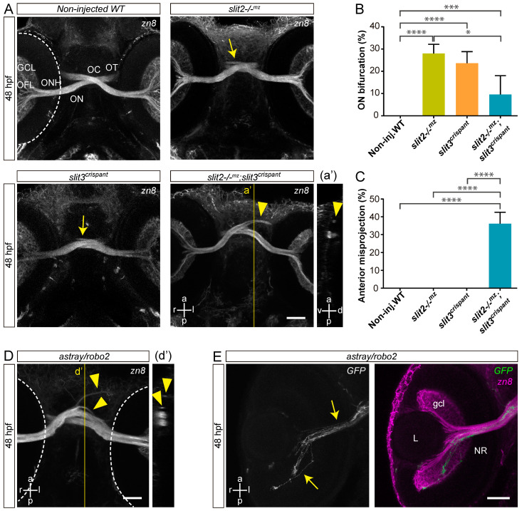
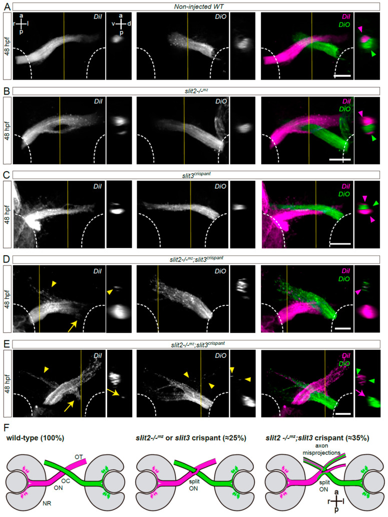
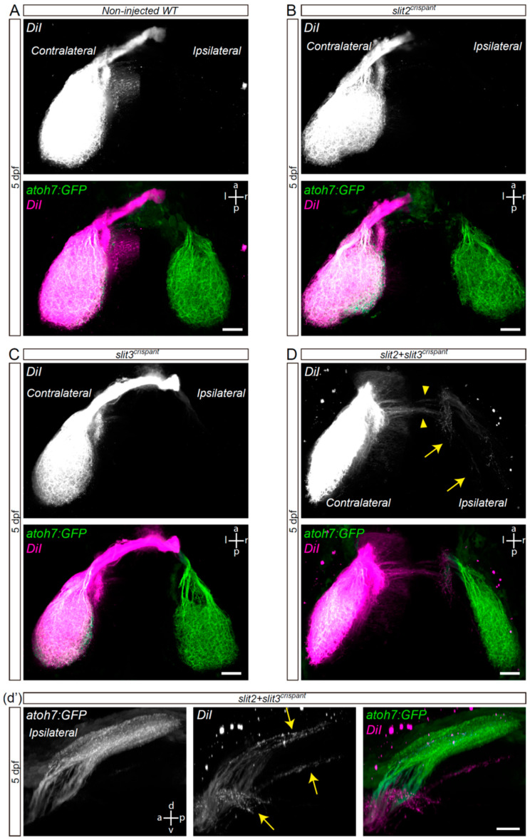
Slit-Robo signaling regulates midline crossing of commissural axons in different systems. In zebrafish, all retinofugal axons cross at the optic chiasm to innervate the contralateral tectum. Here, the mutant for the Robo2 receptor presents severe axon guidance defects, which were not completely reproduced in a Slit2 ligand null mutant. Since slit3 is also expressed around this area at the stage of axon crossing, we decided to analyze the possibility that it collaborates with Slit2 in this process. We found that the disruption of slit3 expression by sgRNA-Cas9 injection caused similar, albeit slightly milder, defects than those of the slit2 mutant, while the same treatment in the slit2-/-mz background caused much more severe defects, comparable to those observed in robo2 mutants. Tracking analysis of in vivo time-lapse experiments indicated differential but complementary functions of these secreted factors in the correction of axon turn errors around the optic chiasm. Interestingly, RT-qPCR analysis showed a mild increase in slit2 expression in slit3-deficient embryos, but not the opposite. Our observations support the previously proposed "repulsive channel" model for Slit-Robo action at the optic chiasm, with both Slits acting in different manners, most probably relating to their different spatial expression patterns.
期刊介绍:
The Journal of Developmental Biology (ISSN 2221-3759) is an international, peer-reviewed, quick-refereeing, open access journal, which publishes reviews, research papers and communications on the development of multicellular organisms at the molecule, cell, tissue, organ and whole organism levels. Our aim is to encourage researchers to effortlessly publish their new findings or concepts rapidly in an open access medium, overseen by their peers. There is no restriction on the length of the papers; the full experimental details must be provided so that the results can be reproduced. Electronic files regarding the full details of the experimental procedure, if unable to be published in a normal way, can be deposited as supplementary material. Journal of Developmental Biology focuses on: -Development mechanisms and genetics -Cell differentiation -Embryonal development -Tissue/organism growth -Metamorphosis and regeneration of the organisms. It involves many biological fields, such as Molecular biology, Genetics, Physiology, Cell biology, Anatomy, Embryology, Cancer research, Neurobiology, Immunology, Ecology, Evolutionary biology.


 分享
分享
 求助内容:
求助内容: 应助结果提醒方式:
 扫码关注我们
