Doaa Abdallah El-Naggar, Laila Mohammed Ahmad El-Zalabany, Doaa Abdelhalim Shahin, Afaf Mahmoud Attia, Shaaban Abdelfattah El-Mosallamy
{"title":"氯二酚对大鼠睾丸的毒性:生化、病理及流式细胞术研究。","authors":"Doaa Abdallah El-Naggar, Laila Mohammed Ahmad El-Zalabany, Doaa Abdelhalim Shahin, Afaf Mahmoud Attia, Shaaban Abdelfattah El-Mosallamy","doi":"10.2147/JEP.S358571","DOIUrl":null,"url":null,"abstract":"<p><strong>Background: </strong>Chloroxylenol (para-chloro-meta-xylenol, PCMX) is claimed to be highly harmful both to humans and the environment. Toxic effects of PCMX on testicular functions are scarcely discussed in the literature.</p><p><strong>Aim of study: </strong>To study testicular toxic effects of PCMX on male Sprague-Dawley rats.</p><p><strong>Materials and methods: </strong>Forty animals were randomly distributed into three groups: negative control (G I), vehicle group (G II) and PCMX group (G III). PCMX group was subdivided into three subgroups: GIIIa: received PCMX 100 mg/kg, GIIIb: received PCMX 200 mg/kg and G IIIc: received PCMX 500 mg/kg. Hormonal assay included assessment of serum testosterone and estradiol levels. Histopathological examination of testicular tissue, analysis of cellular viability, necrosis and apoptosis in testicular tissue by flow cytometry, analysis of cellular DNA content and phases of cell cycle analysis by flow cytometry were also performed.</p><p><strong>Results: </strong>Rats in the groups exposed to PCMX (G IIIa, G IIIb and G IIIc) had significantly lower estradiol and testosterone levels in comparison to control groups (G I and GII). Histopathological examination of testicular tissue of PCMX-exposed rats showed irregular crossly sectioned seminiferous tubules with their lumina containing scanty spermatids and spermatozoa. G IIIc animals showed eosinophilic proteinaceous material and vacuolated and necrotic interstitial cells of Leydig. Rats in PCMX-exposed groups (G IIIa, G IIIb and G IIIc) showed significantly lower testicular tissue viability in comparison to control groups (G I and G II). Rats in PCMX-exposed groups (G IIIa, G IIIb and G IIIc) showed significantly lower percentage of cells in the G0/G1 phase in comparison to control groups (G I and G II).</p><p><strong>Conclusion: </strong>Rats exposed to PCMX had significant reduction in testosterone and estradiol levels with marked histopathological alterations affecting testicular tissues. These effects are dose-dependent.</p>","PeriodicalId":15846,"journal":{"name":"Journal of Experimental Pharmacology","volume":" ","pages":"213-220"},"PeriodicalIF":0.0000,"publicationDate":"2022-07-13","publicationTypes":"Journal Article","fieldsOfStudy":null,"isOpenAccess":false,"openAccessPdf":"https://ftp.ncbi.nlm.nih.gov/pub/pmc/oa_pdf/b0/04/jep-14-213.PMC9289274.pdf","citationCount":"2","resultStr":"{\"title\":\"Testicular Toxicity of Chloroxylenol in Rats: Biochemical, Pathological and Flow Cytometric Study.\",\"authors\":\"Doaa Abdallah El-Naggar, Laila Mohammed Ahmad El-Zalabany, Doaa Abdelhalim Shahin, Afaf Mahmoud Attia, Shaaban Abdelfattah El-Mosallamy\",\"doi\":\"10.2147/JEP.S358571\",\"DOIUrl\":null,\"url\":null,\"abstract\":\"<p><strong>Background: </strong>Chloroxylenol (para-chloro-meta-xylenol, PCMX) is claimed to be highly harmful both to humans and the environment. Toxic effects of PCMX on testicular functions are scarcely discussed in the literature.</p><p><strong>Aim of study: </strong>To study testicular toxic effects of PCMX on male Sprague-Dawley rats.</p><p><strong>Materials and methods: </strong>Forty animals were randomly distributed into three groups: negative control (G I), vehicle group (G II) and PCMX group (G III). PCMX group was subdivided into three subgroups: GIIIa: received PCMX 100 mg/kg, GIIIb: received PCMX 200 mg/kg and G IIIc: received PCMX 500 mg/kg. Hormonal assay included assessment of serum testosterone and estradiol levels. Histopathological examination of testicular tissue, analysis of cellular viability, necrosis and apoptosis in testicular tissue by flow cytometry, analysis of cellular DNA content and phases of cell cycle analysis by flow cytometry were also performed.</p><p><strong>Results: </strong>Rats in the groups exposed to PCMX (G IIIa, G IIIb and G IIIc) had significantly lower estradiol and testosterone levels in comparison to control groups (G I and GII). Histopathological examination of testicular tissue of PCMX-exposed rats showed irregular crossly sectioned seminiferous tubules with their lumina containing scanty spermatids and spermatozoa. G IIIc animals showed eosinophilic proteinaceous material and vacuolated and necrotic interstitial cells of Leydig. Rats in PCMX-exposed groups (G IIIa, G IIIb and G IIIc) showed significantly lower testicular tissue viability in comparison to control groups (G I and G II). Rats in PCMX-exposed groups (G IIIa, G IIIb and G IIIc) showed significantly lower percentage of cells in the G0/G1 phase in comparison to control groups (G I and G II).</p><p><strong>Conclusion: </strong>Rats exposed to PCMX had significant reduction in testosterone and estradiol levels with marked histopathological alterations affecting testicular tissues. These effects are dose-dependent.</p>\",\"PeriodicalId\":15846,\"journal\":{\"name\":\"Journal of Experimental Pharmacology\",\"volume\":\" \",\"pages\":\"213-220\"},\"PeriodicalIF\":0.0000,\"publicationDate\":\"2022-07-13\",\"publicationTypes\":\"Journal Article\",\"fieldsOfStudy\":null,\"isOpenAccess\":false,\"openAccessPdf\":\"https://ftp.ncbi.nlm.nih.gov/pub/pmc/oa_pdf/b0/04/jep-14-213.PMC9289274.pdf\",\"citationCount\":\"2\",\"resultStr\":null,\"platform\":\"Semanticscholar\",\"paperid\":null,\"PeriodicalName\":\"Journal of Experimental Pharmacology\",\"FirstCategoryId\":\"1085\",\"ListUrlMain\":\"https://doi.org/10.2147/JEP.S358571\",\"RegionNum\":0,\"RegionCategory\":null,\"ArticlePicture\":[],\"TitleCN\":null,\"AbstractTextCN\":null,\"PMCID\":null,\"EPubDate\":\"2022/1/1 0:00:00\",\"PubModel\":\"eCollection\",\"JCR\":\"Q2\",\"JCRName\":\"Medicine\",\"Score\":null,\"Total\":0}","platform":"Semanticscholar","paperid":null,"PeriodicalName":"Journal of Experimental Pharmacology","FirstCategoryId":"1085","ListUrlMain":"https://doi.org/10.2147/JEP.S358571","RegionNum":0,"RegionCategory":null,"ArticlePicture":[],"TitleCN":null,"AbstractTextCN":null,"PMCID":null,"EPubDate":"2022/1/1 0:00:00","PubModel":"eCollection","JCR":"Q2","JCRName":"Medicine","Score":null,"Total":0}
Testicular Toxicity of Chloroxylenol in Rats: Biochemical, Pathological and Flow Cytometric Study.
Background: Chloroxylenol (para-chloro-meta-xylenol, PCMX) is claimed to be highly harmful both to humans and the environment. Toxic effects of PCMX on testicular functions are scarcely discussed in the literature.
Aim of study: To study testicular toxic effects of PCMX on male Sprague-Dawley rats.
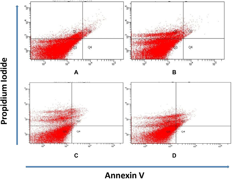
Materials and methods: Forty animals were randomly distributed into three groups: negative control (G I), vehicle group (G II) and PCMX group (G III). PCMX group was subdivided into three subgroups: GIIIa: received PCMX 100 mg/kg, GIIIb: received PCMX 200 mg/kg and G IIIc: received PCMX 500 mg/kg. Hormonal assay included assessment of serum testosterone and estradiol levels. Histopathological examination of testicular tissue, analysis of cellular viability, necrosis and apoptosis in testicular tissue by flow cytometry, analysis of cellular DNA content and phases of cell cycle analysis by flow cytometry were also performed.
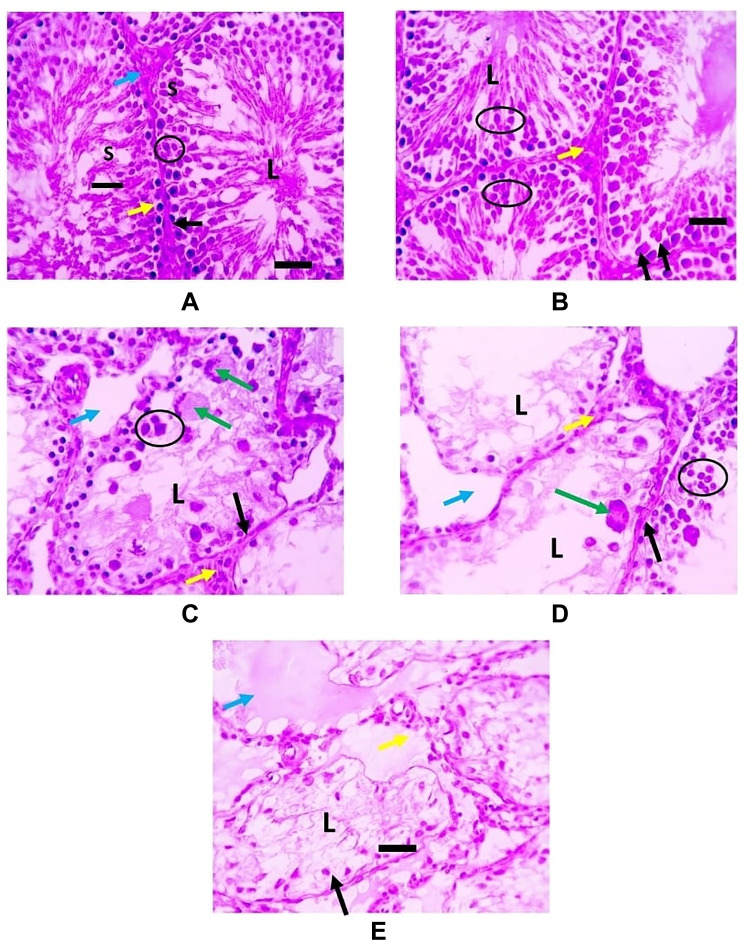
Results: Rats in the groups exposed to PCMX (G IIIa, G IIIb and G IIIc) had significantly lower estradiol and testosterone levels in comparison to control groups (G I and GII). Histopathological examination of testicular tissue of PCMX-exposed rats showed irregular crossly sectioned seminiferous tubules with their lumina containing scanty spermatids and spermatozoa. G IIIc animals showed eosinophilic proteinaceous material and vacuolated and necrotic interstitial cells of Leydig. Rats in PCMX-exposed groups (G IIIa, G IIIb and G IIIc) showed significantly lower testicular tissue viability in comparison to control groups (G I and G II). Rats in PCMX-exposed groups (G IIIa, G IIIb and G IIIc) showed significantly lower percentage of cells in the G0/G1 phase in comparison to control groups (G I and G II).
Conclusion: Rats exposed to PCMX had significant reduction in testosterone and estradiol levels with marked histopathological alterations affecting testicular tissues. These effects are dose-dependent.


 分享
分享
 求助内容:
求助内容: 应助结果提醒方式:
 扫码关注我们
