{"title":"扫描源光学相干断层血管成像视网膜分支静脉闭塞囊性病变的微血管变化。","authors":"Satoko Araki, Susumu Sakimoto, Daiki Shiozaki, Chihiro Ueda, Chikako Hara, Yoko Fukushima, Kaori Sayanagi, Hirokazu Sakaguchi, Kohji Nishida","doi":"10.1159/000525497","DOIUrl":null,"url":null,"abstract":"<p><strong>Introduction: </strong>This study aimed to describe the quantitative features of the microvasculature in the cystic lesions of branch retinal vein occlusion (BRVO).</p><p><strong>Methods: </strong>A total of 43 eyes with BRVO, treated with anti-vascular endothelial growth factor therapy, were analyzed. Using wide-field swept-source optical coherence tomography angiography (OCTA), en face OCT images were obtained by depth-integrated reflectivity of the retina, and vascular density (VD), vascular length (VL), vascular lacunarity, and fractal dimension (FD) were evaluated in a 12 × 12-mm area of retinal nonperfusion.</p><p><strong>Results: </strong>The mean area of affected lesions was 38.7 ± 19.8 mm<sup>2</sup>, and cystic lesions were 8.5 ± 10.1 mm<sup>2</sup>. VD, VL, and FD were significantly decreased in the cystic lesions compared to other affected lesions in the same eyes (<i>p</i> = 0.0010, <i>p</i> = 0.0001, and <i>p</i> = 0.0003, respectively) and in all eyes (<i>p</i> = 0.0281, <i>p</i> = 0.0050, and <i>p</i> < 0.0001, respectively). VD in cystic lesions within the vascular arcade (25 eyes) correlated with best-corrected visual acuity on OCTA (<i>r</i> = -0.433, and <i>p</i> = 0.0492).</p><p><strong>Conclusions: </strong>Vascular structure in the cystic lesions was unpreserved compared to the other lesions in BRVO. These findings may help in understanding the pathophysiology of retinal edema in BRVO.</p>","PeriodicalId":9075,"journal":{"name":"Biomedicine Hub","volume":null,"pages":null},"PeriodicalIF":0.0000,"publicationDate":"2022-08-16","publicationTypes":"Journal Article","fieldsOfStudy":null,"isOpenAccess":false,"openAccessPdf":"https://ftp.ncbi.nlm.nih.gov/pub/pmc/oa_pdf/23/ce/bmh-0007-0099.PMC9574207.pdf","citationCount":"1","resultStr":"{\"title\":\"Microvascular Changes in the Cystic Lesion of Branch Retinal Vein Occlusion Imaged by Swept-Source Optical Coherence Tomography Angiography.\",\"authors\":\"Satoko Araki, Susumu Sakimoto, Daiki Shiozaki, Chihiro Ueda, Chikako Hara, Yoko Fukushima, Kaori Sayanagi, Hirokazu Sakaguchi, Kohji Nishida\",\"doi\":\"10.1159/000525497\",\"DOIUrl\":null,\"url\":null,\"abstract\":\"<p><strong>Introduction: </strong>This study aimed to describe the quantitative features of the microvasculature in the cystic lesions of branch retinal vein occlusion (BRVO).</p><p><strong>Methods: </strong>A total of 43 eyes with BRVO, treated with anti-vascular endothelial growth factor therapy, were analyzed. Using wide-field swept-source optical coherence tomography angiography (OCTA), en face OCT images were obtained by depth-integrated reflectivity of the retina, and vascular density (VD), vascular length (VL), vascular lacunarity, and fractal dimension (FD) were evaluated in a 12 × 12-mm area of retinal nonperfusion.</p><p><strong>Results: </strong>The mean area of affected lesions was 38.7 ± 19.8 mm<sup>2</sup>, and cystic lesions were 8.5 ± 10.1 mm<sup>2</sup>. VD, VL, and FD were significantly decreased in the cystic lesions compared to other affected lesions in the same eyes (<i>p</i> = 0.0010, <i>p</i> = 0.0001, and <i>p</i> = 0.0003, respectively) and in all eyes (<i>p</i> = 0.0281, <i>p</i> = 0.0050, and <i>p</i> < 0.0001, respectively). VD in cystic lesions within the vascular arcade (25 eyes) correlated with best-corrected visual acuity on OCTA (<i>r</i> = -0.433, and <i>p</i> = 0.0492).</p><p><strong>Conclusions: </strong>Vascular structure in the cystic lesions was unpreserved compared to the other lesions in BRVO. These findings may help in understanding the pathophysiology of retinal edema in BRVO.</p>\",\"PeriodicalId\":9075,\"journal\":{\"name\":\"Biomedicine Hub\",\"volume\":null,\"pages\":null},\"PeriodicalIF\":0.0000,\"publicationDate\":\"2022-08-16\",\"publicationTypes\":\"Journal Article\",\"fieldsOfStudy\":null,\"isOpenAccess\":false,\"openAccessPdf\":\"https://ftp.ncbi.nlm.nih.gov/pub/pmc/oa_pdf/23/ce/bmh-0007-0099.PMC9574207.pdf\",\"citationCount\":\"1\",\"resultStr\":null,\"platform\":\"Semanticscholar\",\"paperid\":null,\"PeriodicalName\":\"Biomedicine Hub\",\"FirstCategoryId\":\"1085\",\"ListUrlMain\":\"https://doi.org/10.1159/000525497\",\"RegionNum\":0,\"RegionCategory\":null,\"ArticlePicture\":[],\"TitleCN\":null,\"AbstractTextCN\":null,\"PMCID\":null,\"EPubDate\":\"2022/5/1 0:00:00\",\"PubModel\":\"eCollection\",\"JCR\":\"\",\"JCRName\":\"\",\"Score\":null,\"Total\":0}","platform":"Semanticscholar","paperid":null,"PeriodicalName":"Biomedicine Hub","FirstCategoryId":"1085","ListUrlMain":"https://doi.org/10.1159/000525497","RegionNum":0,"RegionCategory":null,"ArticlePicture":[],"TitleCN":null,"AbstractTextCN":null,"PMCID":null,"EPubDate":"2022/5/1 0:00:00","PubModel":"eCollection","JCR":"","JCRName":"","Score":null,"Total":0}
Microvascular Changes in the Cystic Lesion of Branch Retinal Vein Occlusion Imaged by Swept-Source Optical Coherence Tomography Angiography.
Introduction: This study aimed to describe the quantitative features of the microvasculature in the cystic lesions of branch retinal vein occlusion (BRVO).
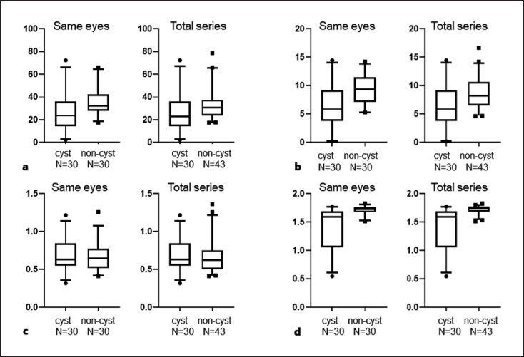
Methods: A total of 43 eyes with BRVO, treated with anti-vascular endothelial growth factor therapy, were analyzed. Using wide-field swept-source optical coherence tomography angiography (OCTA), en face OCT images were obtained by depth-integrated reflectivity of the retina, and vascular density (VD), vascular length (VL), vascular lacunarity, and fractal dimension (FD) were evaluated in a 12 × 12-mm area of retinal nonperfusion.
Results: The mean area of affected lesions was 38.7 ± 19.8 mm2, and cystic lesions were 8.5 ± 10.1 mm2. VD, VL, and FD were significantly decreased in the cystic lesions compared to other affected lesions in the same eyes (p = 0.0010, p = 0.0001, and p = 0.0003, respectively) and in all eyes (p = 0.0281, p = 0.0050, and p < 0.0001, respectively). VD in cystic lesions within the vascular arcade (25 eyes) correlated with best-corrected visual acuity on OCTA (r = -0.433, and p = 0.0492).
Conclusions: Vascular structure in the cystic lesions was unpreserved compared to the other lesions in BRVO. These findings may help in understanding the pathophysiology of retinal edema in BRVO.


 分享
分享

 求助内容:
求助内容: 应助结果提醒方式:
 扫码关注我们
