Hasan M Sharhan, Abeer A Almashraqi, Hanan Al-Fakeh, Najah Alhashimi, Ehab A Abdulghani, Wenyuanfeng Chen, Abeer A Al-Sosowa, BaoCheng Cao, Maged S Alhammadi
{"title":"有上颌阻生牙和没有上颌阻生牙的患者上颌基牙和牙槽牙尺寸的定性和定量三维评价。","authors":"Hasan M Sharhan, Abeer A Almashraqi, Hanan Al-Fakeh, Najah Alhashimi, Ehab A Abdulghani, Wenyuanfeng Chen, Abeer A Al-Sosowa, BaoCheng Cao, Maged S Alhammadi","doi":"10.1186/s40510-022-00434-3","DOIUrl":null,"url":null,"abstract":"<p><strong>Background: </strong>This study aimed to three-dimensionally evaluate the qualitative and quantitative maxillary basal, dentoalveolar, and dental dimensions in patients with unilateral or bilateral maxillary impacted canines relative to their normal peers.</p><p><strong>Materials and methods: </strong>This is a retrospective comparative study. Cone-beam computed tomography images of one hundred and fifty adult patients were divided into three equal groups: unilateral, bilateral, and control groups. Each had 50 patients that were three-dimensionally analysed. The quantitative measurements involved three basal (molar basal width, premolar basal width, and arch depth), seven dentoalveolar (molar alveolar width, premolar alveolar width, inter-molar width, inter-premolar width, inter-canine width, arch length, and arch perimeter), and two dental (canine length and width) measurements. The qualitative measurements included four bone density areas (buccal, lingual, mesial, and distal) around the maxillary impacted canines.</p><p><strong>Result: </strong>Differences between the three groups were statistically different for the quantitative measurements involving the two basal variables (molar basal width and premolar basal width) and all measured dentoalveolar variables; these were smaller in the unilateral and bilateral groups compared with the control group (p < 0.001). Unilateral and bilateral impacted canine groups showed significantly wider and shorter canines than the control group (p < 0.001). The qualitative measurements (the four bone density areas) around unilateral and bilateral impacted canine groups showed significantly greater density than the control group (p < 0.001). There was no significant qualitative or quantitative difference between the unilateral and bilateral impacted canines. The three groups had no significant variations in terms of arch depth.</p><p><strong>Conclusion: </strong>Maxillary unilateral and bilateral canine impactions are associated with reduced basal and dentoalveolar dimensions as well as wider and shorter maxillary canines compared to normal peers. The quality of bone around unilateral and bilateral impacted maxillary canines is higher than in non-impacted cases. Unilateral and bilateral canine impactions have quite similar qualitative and quantitative parameters.</p>","PeriodicalId":56071,"journal":{"name":"Progress in Orthodontics","volume":" ","pages":"38"},"PeriodicalIF":5.0000,"publicationDate":"2022-10-24","publicationTypes":"Journal Article","fieldsOfStudy":null,"isOpenAccess":false,"openAccessPdf":"https://www.ncbi.nlm.nih.gov/pmc/articles/PMC9588850/pdf/","citationCount":"3","resultStr":"{\"title\":\"Qualitative and quantitative three-dimensional evaluation of maxillary basal and dentoalveolar dimensions in patients with and without maxillary impacted canines.\",\"authors\":\"Hasan M Sharhan, Abeer A Almashraqi, Hanan Al-Fakeh, Najah Alhashimi, Ehab A Abdulghani, Wenyuanfeng Chen, Abeer A Al-Sosowa, BaoCheng Cao, Maged S Alhammadi\",\"doi\":\"10.1186/s40510-022-00434-3\",\"DOIUrl\":null,\"url\":null,\"abstract\":\"<p><strong>Background: </strong>This study aimed to three-dimensionally evaluate the qualitative and quantitative maxillary basal, dentoalveolar, and dental dimensions in patients with unilateral or bilateral maxillary impacted canines relative to their normal peers.</p><p><strong>Materials and methods: </strong>This is a retrospective comparative study. Cone-beam computed tomography images of one hundred and fifty adult patients were divided into three equal groups: unilateral, bilateral, and control groups. Each had 50 patients that were three-dimensionally analysed. The quantitative measurements involved three basal (molar basal width, premolar basal width, and arch depth), seven dentoalveolar (molar alveolar width, premolar alveolar width, inter-molar width, inter-premolar width, inter-canine width, arch length, and arch perimeter), and two dental (canine length and width) measurements. The qualitative measurements included four bone density areas (buccal, lingual, mesial, and distal) around the maxillary impacted canines.</p><p><strong>Result: </strong>Differences between the three groups were statistically different for the quantitative measurements involving the two basal variables (molar basal width and premolar basal width) and all measured dentoalveolar variables; these were smaller in the unilateral and bilateral groups compared with the control group (p < 0.001). Unilateral and bilateral impacted canine groups showed significantly wider and shorter canines than the control group (p < 0.001). The qualitative measurements (the four bone density areas) around unilateral and bilateral impacted canine groups showed significantly greater density than the control group (p < 0.001). There was no significant qualitative or quantitative difference between the unilateral and bilateral impacted canines. The three groups had no significant variations in terms of arch depth.</p><p><strong>Conclusion: </strong>Maxillary unilateral and bilateral canine impactions are associated with reduced basal and dentoalveolar dimensions as well as wider and shorter maxillary canines compared to normal peers. The quality of bone around unilateral and bilateral impacted maxillary canines is higher than in non-impacted cases. Unilateral and bilateral canine impactions have quite similar qualitative and quantitative parameters.</p>\",\"PeriodicalId\":56071,\"journal\":{\"name\":\"Progress in Orthodontics\",\"volume\":\" \",\"pages\":\"38\"},\"PeriodicalIF\":5.0000,\"publicationDate\":\"2022-10-24\",\"publicationTypes\":\"Journal Article\",\"fieldsOfStudy\":null,\"isOpenAccess\":false,\"openAccessPdf\":\"https://www.ncbi.nlm.nih.gov/pmc/articles/PMC9588850/pdf/\",\"citationCount\":\"3\",\"resultStr\":null,\"platform\":\"Semanticscholar\",\"paperid\":null,\"PeriodicalName\":\"Progress in Orthodontics\",\"FirstCategoryId\":\"3\",\"ListUrlMain\":\"https://doi.org/10.1186/s40510-022-00434-3\",\"RegionNum\":2,\"RegionCategory\":\"医学\",\"ArticlePicture\":[],\"TitleCN\":null,\"AbstractTextCN\":null,\"PMCID\":null,\"EPubDate\":\"\",\"PubModel\":\"\",\"JCR\":\"Q1\",\"JCRName\":\"Dentistry\",\"Score\":null,\"Total\":0}","platform":"Semanticscholar","paperid":null,"PeriodicalName":"Progress in Orthodontics","FirstCategoryId":"3","ListUrlMain":"https://doi.org/10.1186/s40510-022-00434-3","RegionNum":2,"RegionCategory":"医学","ArticlePicture":[],"TitleCN":null,"AbstractTextCN":null,"PMCID":null,"EPubDate":"","PubModel":"","JCR":"Q1","JCRName":"Dentistry","Score":null,"Total":0}
Qualitative and quantitative three-dimensional evaluation of maxillary basal and dentoalveolar dimensions in patients with and without maxillary impacted canines.
Background: This study aimed to three-dimensionally evaluate the qualitative and quantitative maxillary basal, dentoalveolar, and dental dimensions in patients with unilateral or bilateral maxillary impacted canines relative to their normal peers.
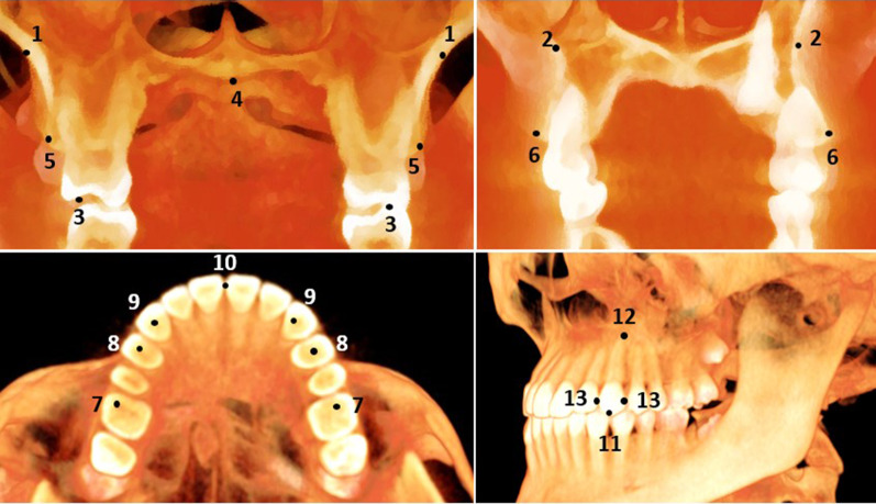
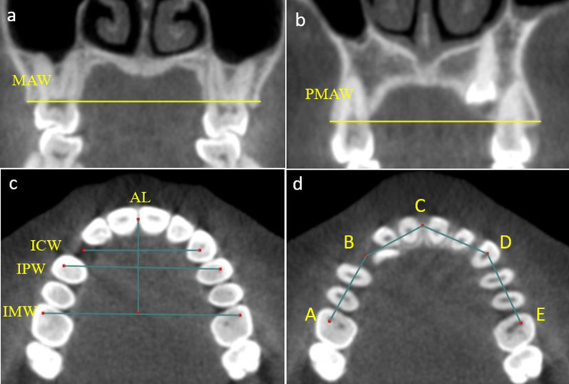
Materials and methods: This is a retrospective comparative study. Cone-beam computed tomography images of one hundred and fifty adult patients were divided into three equal groups: unilateral, bilateral, and control groups. Each had 50 patients that were three-dimensionally analysed. The quantitative measurements involved three basal (molar basal width, premolar basal width, and arch depth), seven dentoalveolar (molar alveolar width, premolar alveolar width, inter-molar width, inter-premolar width, inter-canine width, arch length, and arch perimeter), and two dental (canine length and width) measurements. The qualitative measurements included four bone density areas (buccal, lingual, mesial, and distal) around the maxillary impacted canines.
Result: Differences between the three groups were statistically different for the quantitative measurements involving the two basal variables (molar basal width and premolar basal width) and all measured dentoalveolar variables; these were smaller in the unilateral and bilateral groups compared with the control group (p < 0.001). Unilateral and bilateral impacted canine groups showed significantly wider and shorter canines than the control group (p < 0.001). The qualitative measurements (the four bone density areas) around unilateral and bilateral impacted canine groups showed significantly greater density than the control group (p < 0.001). There was no significant qualitative or quantitative difference between the unilateral and bilateral impacted canines. The three groups had no significant variations in terms of arch depth.
Conclusion: Maxillary unilateral and bilateral canine impactions are associated with reduced basal and dentoalveolar dimensions as well as wider and shorter maxillary canines compared to normal peers. The quality of bone around unilateral and bilateral impacted maxillary canines is higher than in non-impacted cases. Unilateral and bilateral canine impactions have quite similar qualitative and quantitative parameters.
期刊介绍:
Progress in Orthodontics is a fully open access, international journal owned by the Italian Society of Orthodontics and published under the brand SpringerOpen. The Society is currently covering all publication costs so there are no article processing charges for authors.
It is a premier journal of international scope that fosters orthodontic research, including both basic research and development of innovative clinical techniques, with an emphasis on the following areas:
• Mechanisms to improve orthodontics
• Clinical studies and control animal studies
• Orthodontics and genetics, genomics
• Temporomandibular joint (TMJ) control clinical trials
• Efficacy of orthodontic appliances and animal models
• Systematic reviews and meta analyses
• Mechanisms to speed orthodontic treatment
Progress in Orthodontics will consider for publication only meritorious and original contributions. These may be:
• Original articles reporting the findings of clinical trials, clinically relevant basic scientific investigations, or novel therapeutic or diagnostic systems
• Review articles on current topics
• Articles on novel techniques and clinical tools
• Articles of contemporary interest


 分享
分享
 求助内容:
求助内容: 应助结果提醒方式:
 扫码关注我们
