Lech Sedlak, Marta Świerczyńska, Dorota Pojda-Wilczek
{"title":"25岁女性视网膜中央静脉和纤毛视网膜动脉合并闭塞。","authors":"Lech Sedlak, Marta Świerczyńska, Dorota Pojda-Wilczek","doi":"10.22336/rjo.2022.35","DOIUrl":null,"url":null,"abstract":"<p><p>We report a case of a 25-year-old woman with sudden and painless diminution in vision and central scotoma in her left eye (LE). She was a smoker and had been taking combined oral contraceptive (COC) pills for 1 year. On admission, the best-corrected visual acuity (BCVA) was 1,5/50 in the LE. Posterior segment examination revealed optic disc edema with flame-shaped retinal hemorrhages, mildly tortuous and dilated retinal veins. Moreover, retinal edema in the peripapillary and perimacular region, foci of hemorrhages and Roth's spots in the posterior pole, as well as pale superior papillomacular bundle were observed. Fundus fluorescein angiography (FFA) confirmed the delayed flow of contrast through the cilioretinal artery in the LE. The clinical picture suggested left central retinal vein (CRVO) with cilioretinal artery occlusion (CLRAO). All laboratory and imaging tests were normal except for homozygous methylenetetrahydrofolate reductase (MTHFR) gene mutation (A1298C genotypes). However, serum homocysteine (Hcy) level was normal. Low molecular weight heparin (LMWH) treatment was administered. Retinal lesions, as well as BCVA improved, but central scotoma remained. <b>Abbreviations:</b> aPTT = activated partial thromboplastin time, BCVA = best-corrected visual acuity, CBC = complete blood count, CLRAO = cilioretinal artery occlusion, COC = combined oral contraceptive, CRA = central retinal artery, CRP = serum C-reactive protein, CRVO = central retinal vein occlusion, CT = computed tomography, CTA = computed tomography angiography, ECG = electrocardiography, ESR = erythrocyte sedimentation rate, FERG = flash electroretinogram, FFA = fundus fluorescein angiography, GCA = ganglion cell analysis, GCL = ganglion cell layer, Hcy = homocysteine, ICGA = indocyanine green angiography, INR = international normalized ratio, IOP = intraocular pressure, IPL = inner plexiform layer, LE = left eye, LMWH = low molecular weight heparin, mfERG = multifocal electroretinogram, MTHFR = methylenetetrahydrofolate reductase, OCT = optical coherence tomography, RE = right eye, VF = visual field.</p>","PeriodicalId":21385,"journal":{"name":"Romanian journal of ophthalmology","volume":"66 2","pages":"178-184"},"PeriodicalIF":0.0000,"publicationDate":"2022-04-01","publicationTypes":"Journal Article","fieldsOfStudy":null,"isOpenAccess":false,"openAccessPdf":"https://www.ncbi.nlm.nih.gov/pmc/articles/PMC9289772/pdf/","citationCount":"3","resultStr":"{\"title\":\"Combined central retinal vein and cilioretinal artery occlusion in a 25-year-old woman.\",\"authors\":\"Lech Sedlak, Marta Świerczyńska, Dorota Pojda-Wilczek\",\"doi\":\"10.22336/rjo.2022.35\",\"DOIUrl\":null,\"url\":null,\"abstract\":\"<p><p>We report a case of a 25-year-old woman with sudden and painless diminution in vision and central scotoma in her left eye (LE). She was a smoker and had been taking combined oral contraceptive (COC) pills for 1 year. On admission, the best-corrected visual acuity (BCVA) was 1,5/50 in the LE. Posterior segment examination revealed optic disc edema with flame-shaped retinal hemorrhages, mildly tortuous and dilated retinal veins. Moreover, retinal edema in the peripapillary and perimacular region, foci of hemorrhages and Roth's spots in the posterior pole, as well as pale superior papillomacular bundle were observed. Fundus fluorescein angiography (FFA) confirmed the delayed flow of contrast through the cilioretinal artery in the LE. The clinical picture suggested left central retinal vein (CRVO) with cilioretinal artery occlusion (CLRAO). All laboratory and imaging tests were normal except for homozygous methylenetetrahydrofolate reductase (MTHFR) gene mutation (A1298C genotypes). However, serum homocysteine (Hcy) level was normal. Low molecular weight heparin (LMWH) treatment was administered. Retinal lesions, as well as BCVA improved, but central scotoma remained. <b>Abbreviations:</b> aPTT = activated partial thromboplastin time, BCVA = best-corrected visual acuity, CBC = complete blood count, CLRAO = cilioretinal artery occlusion, COC = combined oral contraceptive, CRA = central retinal artery, CRP = serum C-reactive protein, CRVO = central retinal vein occlusion, CT = computed tomography, CTA = computed tomography angiography, ECG = electrocardiography, ESR = erythrocyte sedimentation rate, FERG = flash electroretinogram, FFA = fundus fluorescein angiography, GCA = ganglion cell analysis, GCL = ganglion cell layer, Hcy = homocysteine, ICGA = indocyanine green angiography, INR = international normalized ratio, IOP = intraocular pressure, IPL = inner plexiform layer, LE = left eye, LMWH = low molecular weight heparin, mfERG = multifocal electroretinogram, MTHFR = methylenetetrahydrofolate reductase, OCT = optical coherence tomography, RE = right eye, VF = visual field.</p>\",\"PeriodicalId\":21385,\"journal\":{\"name\":\"Romanian journal of ophthalmology\",\"volume\":\"66 2\",\"pages\":\"178-184\"},\"PeriodicalIF\":0.0000,\"publicationDate\":\"2022-04-01\",\"publicationTypes\":\"Journal Article\",\"fieldsOfStudy\":null,\"isOpenAccess\":false,\"openAccessPdf\":\"https://www.ncbi.nlm.nih.gov/pmc/articles/PMC9289772/pdf/\",\"citationCount\":\"3\",\"resultStr\":null,\"platform\":\"Semanticscholar\",\"paperid\":null,\"PeriodicalName\":\"Romanian journal of ophthalmology\",\"FirstCategoryId\":\"1085\",\"ListUrlMain\":\"https://doi.org/10.22336/rjo.2022.35\",\"RegionNum\":0,\"RegionCategory\":null,\"ArticlePicture\":[],\"TitleCN\":null,\"AbstractTextCN\":null,\"PMCID\":null,\"EPubDate\":\"\",\"PubModel\":\"\",\"JCR\":\"\",\"JCRName\":\"\",\"Score\":null,\"Total\":0}","platform":"Semanticscholar","paperid":null,"PeriodicalName":"Romanian journal of ophthalmology","FirstCategoryId":"1085","ListUrlMain":"https://doi.org/10.22336/rjo.2022.35","RegionNum":0,"RegionCategory":null,"ArticlePicture":[],"TitleCN":null,"AbstractTextCN":null,"PMCID":null,"EPubDate":"","PubModel":"","JCR":"","JCRName":"","Score":null,"Total":0}
Combined central retinal vein and cilioretinal artery occlusion in a 25-year-old woman.
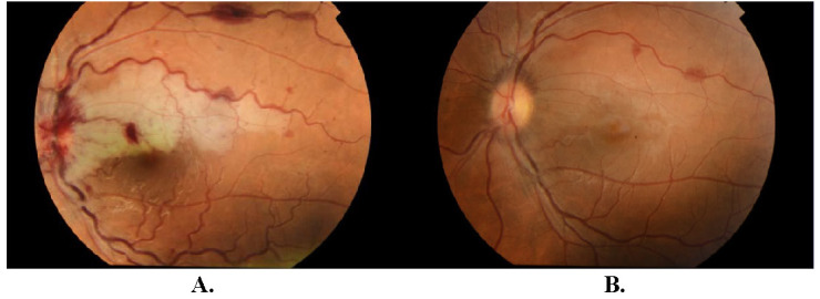
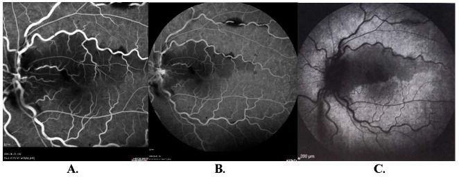
We report a case of a 25-year-old woman with sudden and painless diminution in vision and central scotoma in her left eye (LE). She was a smoker and had been taking combined oral contraceptive (COC) pills for 1 year. On admission, the best-corrected visual acuity (BCVA) was 1,5/50 in the LE. Posterior segment examination revealed optic disc edema with flame-shaped retinal hemorrhages, mildly tortuous and dilated retinal veins. Moreover, retinal edema in the peripapillary and perimacular region, foci of hemorrhages and Roth's spots in the posterior pole, as well as pale superior papillomacular bundle were observed. Fundus fluorescein angiography (FFA) confirmed the delayed flow of contrast through the cilioretinal artery in the LE. The clinical picture suggested left central retinal vein (CRVO) with cilioretinal artery occlusion (CLRAO). All laboratory and imaging tests were normal except for homozygous methylenetetrahydrofolate reductase (MTHFR) gene mutation (A1298C genotypes). However, serum homocysteine (Hcy) level was normal. Low molecular weight heparin (LMWH) treatment was administered. Retinal lesions, as well as BCVA improved, but central scotoma remained. Abbreviations: aPTT = activated partial thromboplastin time, BCVA = best-corrected visual acuity, CBC = complete blood count, CLRAO = cilioretinal artery occlusion, COC = combined oral contraceptive, CRA = central retinal artery, CRP = serum C-reactive protein, CRVO = central retinal vein occlusion, CT = computed tomography, CTA = computed tomography angiography, ECG = electrocardiography, ESR = erythrocyte sedimentation rate, FERG = flash electroretinogram, FFA = fundus fluorescein angiography, GCA = ganglion cell analysis, GCL = ganglion cell layer, Hcy = homocysteine, ICGA = indocyanine green angiography, INR = international normalized ratio, IOP = intraocular pressure, IPL = inner plexiform layer, LE = left eye, LMWH = low molecular weight heparin, mfERG = multifocal electroretinogram, MTHFR = methylenetetrahydrofolate reductase, OCT = optical coherence tomography, RE = right eye, VF = visual field.


 分享
分享

 求助内容:
求助内容: 应助结果提醒方式:
 扫码关注我们
