Da Woon Lee, Si Hyun Kwak, Hwan Jun Choi, Jun Hyuk Kim
{"title":"应用克氏针及眉部小切口复位额窦前壁粉碎性骨折。","authors":"Da Woon Lee, Si Hyun Kwak, Hwan Jun Choi, Jun Hyuk Kim","doi":"10.7181/acfs.2022.00934","DOIUrl":null,"url":null,"abstract":"<p><strong>Background: </strong>Frontal sinus fractures are relatively rare. Their surgical management significantly differs depending on whether the posterior wall is invaded and the clinical features vary. A bicoronal incision or endoscopic approach can be used. However, the minimally invasive approach has been attracting attention, leading us to introduce a simple and effective surgical method using multiple-threaded Kirschner wires.</p><p><strong>Methods: </strong>All patients had isolated anterior wall fractures without nasofrontal duct impairment. The depth from the skin to the posterior wall was measured using computed tomography to prevent injury. The edge of the bone segment on the skin was marked, a threaded Kirschner wire was inserted into the center of the bone segment, and multiple Kirschner wires were gently reduced simultaneously.</p><p><strong>Results: </strong>Surgery was performed on 11 patients. Among them, seven patients required additional support for appropriate fracture reduction. Therefore, a periosteal elevator was used as an adjunct through a small sub-brow incision because the reduction was incomplete with the Kirschner wire alone. The reduction results were confirmed using facial bone computed tomography 1 to 3 days postoperatively. The follow-up period was 3 to 12 months.</p><p><strong>Conclusion: </strong>The patients had no complications and were satisfied with the surgical results. Here we demonstrated an easy and successful procedure to reduce a pure anterior wall frontal sinus fracture via non-invasive threaded Kirschner wire reduction.</p>","PeriodicalId":52238,"journal":{"name":"Archives of Craniofacial Surgery","volume":"23 5","pages":"220-227"},"PeriodicalIF":0.0000,"publicationDate":"2022-10-01","publicationTypes":"Journal Article","fieldsOfStudy":null,"isOpenAccess":false,"openAccessPdf":"https://ftp.ncbi.nlm.nih.gov/pub/pmc/oa_pdf/8c/94/acfs-2022-00934.PMC9663263.pdf","citationCount":"0","resultStr":"{\"title\":\"Reduction of comminuted fractures of the anterior wall of the frontal sinus using threaded Kirschner wires and a small eyebrow incision.\",\"authors\":\"Da Woon Lee, Si Hyun Kwak, Hwan Jun Choi, Jun Hyuk Kim\",\"doi\":\"10.7181/acfs.2022.00934\",\"DOIUrl\":null,\"url\":null,\"abstract\":\"<p><strong>Background: </strong>Frontal sinus fractures are relatively rare. Their surgical management significantly differs depending on whether the posterior wall is invaded and the clinical features vary. A bicoronal incision or endoscopic approach can be used. However, the minimally invasive approach has been attracting attention, leading us to introduce a simple and effective surgical method using multiple-threaded Kirschner wires.</p><p><strong>Methods: </strong>All patients had isolated anterior wall fractures without nasofrontal duct impairment. The depth from the skin to the posterior wall was measured using computed tomography to prevent injury. The edge of the bone segment on the skin was marked, a threaded Kirschner wire was inserted into the center of the bone segment, and multiple Kirschner wires were gently reduced simultaneously.</p><p><strong>Results: </strong>Surgery was performed on 11 patients. Among them, seven patients required additional support for appropriate fracture reduction. Therefore, a periosteal elevator was used as an adjunct through a small sub-brow incision because the reduction was incomplete with the Kirschner wire alone. The reduction results were confirmed using facial bone computed tomography 1 to 3 days postoperatively. The follow-up period was 3 to 12 months.</p><p><strong>Conclusion: </strong>The patients had no complications and were satisfied with the surgical results. Here we demonstrated an easy and successful procedure to reduce a pure anterior wall frontal sinus fracture via non-invasive threaded Kirschner wire reduction.</p>\",\"PeriodicalId\":52238,\"journal\":{\"name\":\"Archives of Craniofacial Surgery\",\"volume\":\"23 5\",\"pages\":\"220-227\"},\"PeriodicalIF\":0.0000,\"publicationDate\":\"2022-10-01\",\"publicationTypes\":\"Journal Article\",\"fieldsOfStudy\":null,\"isOpenAccess\":false,\"openAccessPdf\":\"https://ftp.ncbi.nlm.nih.gov/pub/pmc/oa_pdf/8c/94/acfs-2022-00934.PMC9663263.pdf\",\"citationCount\":\"0\",\"resultStr\":null,\"platform\":\"Semanticscholar\",\"paperid\":null,\"PeriodicalName\":\"Archives of Craniofacial Surgery\",\"FirstCategoryId\":\"1085\",\"ListUrlMain\":\"https://doi.org/10.7181/acfs.2022.00934\",\"RegionNum\":0,\"RegionCategory\":null,\"ArticlePicture\":[],\"TitleCN\":null,\"AbstractTextCN\":null,\"PMCID\":null,\"EPubDate\":\"2022/10/20 0:00:00\",\"PubModel\":\"Epub\",\"JCR\":\"Q2\",\"JCRName\":\"Medicine\",\"Score\":null,\"Total\":0}","platform":"Semanticscholar","paperid":null,"PeriodicalName":"Archives of Craniofacial Surgery","FirstCategoryId":"1085","ListUrlMain":"https://doi.org/10.7181/acfs.2022.00934","RegionNum":0,"RegionCategory":null,"ArticlePicture":[],"TitleCN":null,"AbstractTextCN":null,"PMCID":null,"EPubDate":"2022/10/20 0:00:00","PubModel":"Epub","JCR":"Q2","JCRName":"Medicine","Score":null,"Total":0}
Reduction of comminuted fractures of the anterior wall of the frontal sinus using threaded Kirschner wires and a small eyebrow incision.
Background: Frontal sinus fractures are relatively rare. Their surgical management significantly differs depending on whether the posterior wall is invaded and the clinical features vary. A bicoronal incision or endoscopic approach can be used. However, the minimally invasive approach has been attracting attention, leading us to introduce a simple and effective surgical method using multiple-threaded Kirschner wires.
Methods: All patients had isolated anterior wall fractures without nasofrontal duct impairment. The depth from the skin to the posterior wall was measured using computed tomography to prevent injury. The edge of the bone segment on the skin was marked, a threaded Kirschner wire was inserted into the center of the bone segment, and multiple Kirschner wires were gently reduced simultaneously.
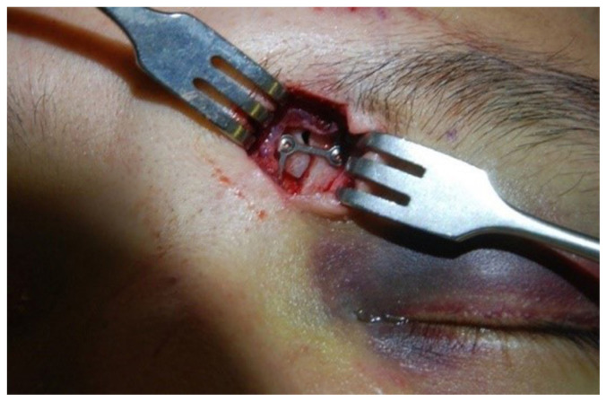
Results: Surgery was performed on 11 patients. Among them, seven patients required additional support for appropriate fracture reduction. Therefore, a periosteal elevator was used as an adjunct through a small sub-brow incision because the reduction was incomplete with the Kirschner wire alone. The reduction results were confirmed using facial bone computed tomography 1 to 3 days postoperatively. The follow-up period was 3 to 12 months.
Conclusion: The patients had no complications and were satisfied with the surgical results. Here we demonstrated an easy and successful procedure to reduce a pure anterior wall frontal sinus fracture via non-invasive threaded Kirschner wire reduction.


 分享
分享
 求助内容:
求助内容: 应助结果提醒方式:
 扫码关注我们
