下载PDF
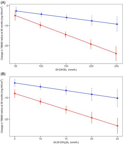
{"title":"卡尔加里维生素D研究中维生素D代谢组的测量:维生素D代谢产物与骨丢失的关系","authors":"Lauren A. Burt, Martin Kaufmann, Marianne S. Rose, Glenville Jones, Emma O. Billington, Steven K. Boyd, David A. Hanley","doi":"10.1002/jbmr.4876","DOIUrl":null,"url":null,"abstract":"<p>In a 36-month randomized controlled trial examining the effect of high-dose vitamin D<sub>3</sub> on radial and tibial total bone mineral density (TtBMD), measured by high-resolution peripheral quantitative tomography (HR-pQCT), participants (311 healthy males and females aged 55–70 years with dual-energy X-ray absorptiometry T-scores > −2.5 without vitamin D deficiency) were randomized to receive 400 IU (<i>N</i> = 109), 4000 IU (<i>N</i> = 100), or 10,000 IU (<i>N</i> = 102) daily. Participants had HR-pQCT radius and tibia scans and blood sampling at baseline, 6, 12, 24, and 36 months. This secondary analysis examined the effect of vitamin D dose on plasma measurements of the vitamin D metabolome by liquid chromatography–tandem mass spectrometry (LC-MS/MS), exploring whether the observed decline in TtBMD was associated with changes in four key metabolites [25-(OH)D<sub>3</sub>; 24,25-(OH)<sub>2</sub>D<sub>3</sub>; 1,25-(OH)<sub>2</sub>D<sub>3</sub>; and 1,24,25-(OH)<sub>3</sub>D<sub>3</sub>]. The relationship between peak values in vitamin D metabolites and changes in TtBMD over 36 months was assessed using linear regression, controlling for sex. Increasing vitamin D dose was associated with a marked increase in 25-(OH)D<sub>3</sub>, 24,25-(OH)<sub>2</sub>D<sub>3</sub> and 1,24,25-(OH)<sub>3</sub>D<sub>3</sub>, but no dose-related change in plasma 1,25-(OH)<sub>2</sub>D<sub>3</sub> was observed. There was a significant negative slope for radius TtBMD and 1,24,25-(OH)<sub>3</sub>D<sub>3</sub> (−0.05, 95% confidence interval [CI] −0.08, −0.03, <i>p</i> < 0.001) after controlling for sex. A significant interaction between TtBMD and sex was seen for 25-(OH)D<sub>3</sub> (female: −0.01, 95% CI −0.12, −0.07; male: −0.04, 95% CI −0.06, −0.01, <i>p</i> = 0.001) and 24,25-(OH)<sub>2</sub>D<sub>3</sub> (female: −0.75, 95% CI −0.98, −0.52; male: −0.35, 95% CI −0.59, −0.11, <i>p</i> < 0.001). For the tibia there was a significant negative slope for 25-(OH)D<sub>3</sub> (−0.03, 95% CI −0.05, −0.01, <i>p</i> < 0.001), 24,25-(OH)<sub>2</sub>D<sub>3</sub> (−0.30, 95% CI −0.44, −0.16, <i>p</i> < 0.001), and 1,24,25-(OH)<sub>3</sub>D<sub>3</sub> (−0.03, 95% CI −0.05, −0.01, <i>p</i> = 0.01) after controlling for sex. These results suggest vitamin D metabolites other than 1,25-(OH)<sub>2</sub>D<sub>3</sub> may be responsible for the bone loss seen in the Calgary Vitamin D Study. Although plasma 1,25-(OH)<sub>2</sub>D<sub>3</sub> did not change with vitamin D dose, it is possible rapid catabolism to 1,24,25-(OH)<sub>3</sub>D<sub>3</sub> prevented the detection of a dose-related rise in plasma 1,25-(OH)<sub>2</sub>D<sub>3</sub>. © 2023 The Authors. <i>Journal of Bone and Mineral Research</i> published by Wiley Periodicals LLC on behalf of American Society for Bone and Mineral Research (ASBMR).</p>","PeriodicalId":185,"journal":{"name":"Journal of Bone and Mineral Research","volume":"38 9","pages":"1312-1321"},"PeriodicalIF":5.1000,"publicationDate":"2023-07-06","publicationTypes":"Journal Article","fieldsOfStudy":null,"isOpenAccess":false,"openAccessPdf":"https://onlinelibrary.wiley.com/doi/epdf/10.1002/jbmr.4876","citationCount":"0","resultStr":"{\"title\":\"Measurements of the Vitamin D Metabolome in the Calgary Vitamin D Study: Relationship of Vitamin D Metabolites to Bone Loss\",\"authors\":\"Lauren A. Burt, Martin Kaufmann, Marianne S. Rose, Glenville Jones, Emma O. Billington, Steven K. Boyd, David A. Hanley\",\"doi\":\"10.1002/jbmr.4876\",\"DOIUrl\":null,\"url\":null,\"abstract\":\"<p>In a 36-month randomized controlled trial examining the effect of high-dose vitamin D<sub>3</sub> on radial and tibial total bone mineral density (TtBMD), measured by high-resolution peripheral quantitative tomography (HR-pQCT), participants (311 healthy males and females aged 55–70 years with dual-energy X-ray absorptiometry T-scores > −2.5 without vitamin D deficiency) were randomized to receive 400 IU (<i>N</i> = 109), 4000 IU (<i>N</i> = 100), or 10,000 IU (<i>N</i> = 102) daily. Participants had HR-pQCT radius and tibia scans and blood sampling at baseline, 6, 12, 24, and 36 months. This secondary analysis examined the effect of vitamin D dose on plasma measurements of the vitamin D metabolome by liquid chromatography–tandem mass spectrometry (LC-MS/MS), exploring whether the observed decline in TtBMD was associated with changes in four key metabolites [25-(OH)D<sub>3</sub>; 24,25-(OH)<sub>2</sub>D<sub>3</sub>; 1,25-(OH)<sub>2</sub>D<sub>3</sub>; and 1,24,25-(OH)<sub>3</sub>D<sub>3</sub>]. The relationship between peak values in vitamin D metabolites and changes in TtBMD over 36 months was assessed using linear regression, controlling for sex. Increasing vitamin D dose was associated with a marked increase in 25-(OH)D<sub>3</sub>, 24,25-(OH)<sub>2</sub>D<sub>3</sub> and 1,24,25-(OH)<sub>3</sub>D<sub>3</sub>, but no dose-related change in plasma 1,25-(OH)<sub>2</sub>D<sub>3</sub> was observed. There was a significant negative slope for radius TtBMD and 1,24,25-(OH)<sub>3</sub>D<sub>3</sub> (−0.05, 95% confidence interval [CI] −0.08, −0.03, <i>p</i> < 0.001) after controlling for sex. A significant interaction between TtBMD and sex was seen for 25-(OH)D<sub>3</sub> (female: −0.01, 95% CI −0.12, −0.07; male: −0.04, 95% CI −0.06, −0.01, <i>p</i> = 0.001) and 24,25-(OH)<sub>2</sub>D<sub>3</sub> (female: −0.75, 95% CI −0.98, −0.52; male: −0.35, 95% CI −0.59, −0.11, <i>p</i> < 0.001). For the tibia there was a significant negative slope for 25-(OH)D<sub>3</sub> (−0.03, 95% CI −0.05, −0.01, <i>p</i> < 0.001), 24,25-(OH)<sub>2</sub>D<sub>3</sub> (−0.30, 95% CI −0.44, −0.16, <i>p</i> < 0.001), and 1,24,25-(OH)<sub>3</sub>D<sub>3</sub> (−0.03, 95% CI −0.05, −0.01, <i>p</i> = 0.01) after controlling for sex. These results suggest vitamin D metabolites other than 1,25-(OH)<sub>2</sub>D<sub>3</sub> may be responsible for the bone loss seen in the Calgary Vitamin D Study. Although plasma 1,25-(OH)<sub>2</sub>D<sub>3</sub> did not change with vitamin D dose, it is possible rapid catabolism to 1,24,25-(OH)<sub>3</sub>D<sub>3</sub> prevented the detection of a dose-related rise in plasma 1,25-(OH)<sub>2</sub>D<sub>3</sub>. © 2023 The Authors. <i>Journal of Bone and Mineral Research</i> published by Wiley Periodicals LLC on behalf of American Society for Bone and Mineral Research (ASBMR).</p>\",\"PeriodicalId\":185,\"journal\":{\"name\":\"Journal of Bone and Mineral Research\",\"volume\":\"38 9\",\"pages\":\"1312-1321\"},\"PeriodicalIF\":5.1000,\"publicationDate\":\"2023-07-06\",\"publicationTypes\":\"Journal Article\",\"fieldsOfStudy\":null,\"isOpenAccess\":false,\"openAccessPdf\":\"https://onlinelibrary.wiley.com/doi/epdf/10.1002/jbmr.4876\",\"citationCount\":\"0\",\"resultStr\":null,\"platform\":\"Semanticscholar\",\"paperid\":null,\"PeriodicalName\":\"Journal of Bone and Mineral Research\",\"FirstCategoryId\":\"3\",\"ListUrlMain\":\"https://onlinelibrary.wiley.com/doi/10.1002/jbmr.4876\",\"RegionNum\":1,\"RegionCategory\":\"医学\",\"ArticlePicture\":[],\"TitleCN\":null,\"AbstractTextCN\":null,\"PMCID\":null,\"EPubDate\":\"\",\"PubModel\":\"\",\"JCR\":\"Q1\",\"JCRName\":\"ENDOCRINOLOGY & METABOLISM\",\"Score\":null,\"Total\":0}","platform":"Semanticscholar","paperid":null,"PeriodicalName":"Journal of Bone and Mineral Research","FirstCategoryId":"3","ListUrlMain":"https://onlinelibrary.wiley.com/doi/10.1002/jbmr.4876","RegionNum":1,"RegionCategory":"医学","ArticlePicture":[],"TitleCN":null,"AbstractTextCN":null,"PMCID":null,"EPubDate":"","PubModel":"","JCR":"Q1","JCRName":"ENDOCRINOLOGY & METABOLISM","Score":null,"Total":0}
引用次数: 0
引用
批量引用


