Surya Prakash Vankina, Surekha Goyal, Geeta S Narayanan
{"title":"癌症放射治疗入口的上限:在目前的3D适形治疗时代,继续使用骨标志是否明智?","authors":"Surya Prakash Vankina, Surekha Goyal, Geeta S Narayanan","doi":"10.5603/RPOR.a2023.0045","DOIUrl":null,"url":null,"abstract":"<p><strong>Background: </strong>This study aimed to compare the levels of L5-S1 interspace and the bifurcation of common iliac vessels on simulation images of rectal cancer patients to evaluate the adequacy of superior borders in conventional 2D planning for covering internal iliac vessels.</p><p><strong>Materials and methods: </strong>Simulation images of 236 rectal cancer patients who received neoadjuvant chemoradiation and surgery were analyzed. The images were retrieved from the radiation treatment database and included delineations of L5-S1 interspace and common iliac vessel bifurcation. Distances between these landmarks were measured.</p><p><strong>Results: </strong>Among the 236 patients, the majority had the common iliac artery bifurcation positioned above the L5-S1 interspace. Specifically, 78.3% of patients had the right common iliac bifurcation above L5-S1 interspace, with an average distance of 2.02 cm. For the left common iliac artery, 77.11% of patients had the bifurcation above L5-S1 interspace, with an average distance of 1.99 cm. Notably, there were cases where the bifurcations were not at the same level.</p><p><strong>Conclusion: </strong>Using the L5-S1 junction as the upper border of the treatment portal may result in missing proximal nodes at risk of metastases. However, further research is needed to determine the significance of failures above the L5-S1 interspace for justifying the inclusion of the common iliac artery bifurcation in the treatment portal.</p>","PeriodicalId":47283,"journal":{"name":"Reports of Practical Oncology and Radiotherapy","volume":"28 4","pages":"565-569"},"PeriodicalIF":1.2000,"publicationDate":"2023-08-28","publicationTypes":"Journal Article","fieldsOfStudy":null,"isOpenAccess":false,"openAccessPdf":"https://ftp.ncbi.nlm.nih.gov/pub/pmc/oa_pdf/54/0d/rpor-28-4-565.PMC10547415.pdf","citationCount":"0","resultStr":"{\"title\":\"Upper limit of radiation treatment portals in rectal cancer: is it wise to keep using bony landmarks in the present era of 3D conformal treatment?\",\"authors\":\"Surya Prakash Vankina, Surekha Goyal, Geeta S Narayanan\",\"doi\":\"10.5603/RPOR.a2023.0045\",\"DOIUrl\":null,\"url\":null,\"abstract\":\"<p><strong>Background: </strong>This study aimed to compare the levels of L5-S1 interspace and the bifurcation of common iliac vessels on simulation images of rectal cancer patients to evaluate the adequacy of superior borders in conventional 2D planning for covering internal iliac vessels.</p><p><strong>Materials and methods: </strong>Simulation images of 236 rectal cancer patients who received neoadjuvant chemoradiation and surgery were analyzed. The images were retrieved from the radiation treatment database and included delineations of L5-S1 interspace and common iliac vessel bifurcation. Distances between these landmarks were measured.</p><p><strong>Results: </strong>Among the 236 patients, the majority had the common iliac artery bifurcation positioned above the L5-S1 interspace. Specifically, 78.3% of patients had the right common iliac bifurcation above L5-S1 interspace, with an average distance of 2.02 cm. For the left common iliac artery, 77.11% of patients had the bifurcation above L5-S1 interspace, with an average distance of 1.99 cm. Notably, there were cases where the bifurcations were not at the same level.</p><p><strong>Conclusion: </strong>Using the L5-S1 junction as the upper border of the treatment portal may result in missing proximal nodes at risk of metastases. However, further research is needed to determine the significance of failures above the L5-S1 interspace for justifying the inclusion of the common iliac artery bifurcation in the treatment portal.</p>\",\"PeriodicalId\":47283,\"journal\":{\"name\":\"Reports of Practical Oncology and Radiotherapy\",\"volume\":\"28 4\",\"pages\":\"565-569\"},\"PeriodicalIF\":1.2000,\"publicationDate\":\"2023-08-28\",\"publicationTypes\":\"Journal Article\",\"fieldsOfStudy\":null,\"isOpenAccess\":false,\"openAccessPdf\":\"https://ftp.ncbi.nlm.nih.gov/pub/pmc/oa_pdf/54/0d/rpor-28-4-565.PMC10547415.pdf\",\"citationCount\":\"0\",\"resultStr\":null,\"platform\":\"Semanticscholar\",\"paperid\":null,\"PeriodicalName\":\"Reports of Practical Oncology and Radiotherapy\",\"FirstCategoryId\":\"1085\",\"ListUrlMain\":\"https://doi.org/10.5603/RPOR.a2023.0045\",\"RegionNum\":0,\"RegionCategory\":null,\"ArticlePicture\":[],\"TitleCN\":null,\"AbstractTextCN\":null,\"PMCID\":null,\"EPubDate\":\"2023/1/1 0:00:00\",\"PubModel\":\"eCollection\",\"JCR\":\"Q4\",\"JCRName\":\"ONCOLOGY\",\"Score\":null,\"Total\":0}","platform":"Semanticscholar","paperid":null,"PeriodicalName":"Reports of Practical Oncology and Radiotherapy","FirstCategoryId":"1085","ListUrlMain":"https://doi.org/10.5603/RPOR.a2023.0045","RegionNum":0,"RegionCategory":null,"ArticlePicture":[],"TitleCN":null,"AbstractTextCN":null,"PMCID":null,"EPubDate":"2023/1/1 0:00:00","PubModel":"eCollection","JCR":"Q4","JCRName":"ONCOLOGY","Score":null,"Total":0}
Upper limit of radiation treatment portals in rectal cancer: is it wise to keep using bony landmarks in the present era of 3D conformal treatment?
Background: This study aimed to compare the levels of L5-S1 interspace and the bifurcation of common iliac vessels on simulation images of rectal cancer patients to evaluate the adequacy of superior borders in conventional 2D planning for covering internal iliac vessels.
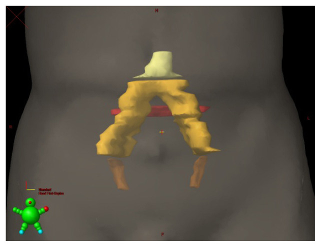
Materials and methods: Simulation images of 236 rectal cancer patients who received neoadjuvant chemoradiation and surgery were analyzed. The images were retrieved from the radiation treatment database and included delineations of L5-S1 interspace and common iliac vessel bifurcation. Distances between these landmarks were measured.
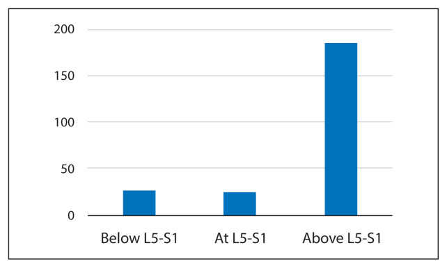
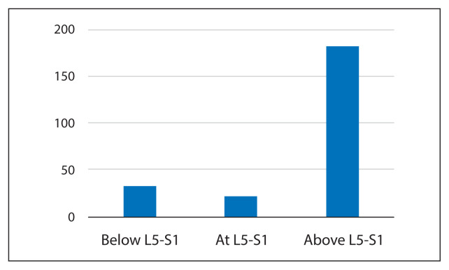
Results: Among the 236 patients, the majority had the common iliac artery bifurcation positioned above the L5-S1 interspace. Specifically, 78.3% of patients had the right common iliac bifurcation above L5-S1 interspace, with an average distance of 2.02 cm. For the left common iliac artery, 77.11% of patients had the bifurcation above L5-S1 interspace, with an average distance of 1.99 cm. Notably, there were cases where the bifurcations were not at the same level.
Conclusion: Using the L5-S1 junction as the upper border of the treatment portal may result in missing proximal nodes at risk of metastases. However, further research is needed to determine the significance of failures above the L5-S1 interspace for justifying the inclusion of the common iliac artery bifurcation in the treatment portal.
期刊介绍:
Reports of Practical Oncology and Radiotherapy is an interdisciplinary bimonthly journal, publishing original contributions in clinical oncology and radiotherapy, as well as in radiotherapy physics, techniques and radiotherapy equipment. Reports of Practical Oncology and Radiotherapy is a journal of the Polish Society of Radiation Oncology, the Czech Society of Radiation Oncology, the Hungarian Society for Radiation Oncology, the Slovenian Society for Radiotherapy and Oncology, the Polish Study Group of Head and Neck Cancer, the Guild of Bulgarian Radiotherapists and the Greater Poland Cancer Centre, affiliated with the Spanish Society of Radiotherapy and Oncology, the Italian Association of Radiotherapy and the Portuguese Society of Radiotherapy - Oncology.


 分享
分享

 求助内容:
求助内容: 应助结果提醒方式:
 扫码关注我们
