Adugna Getachew Mideksa, Tolesa Yadeta Huluka, Masresha Solomon Dino, Mensur Mohammed Ahmed
{"title":"圣保罗医院千禧医学院治疗腔静脉后输尿管的经验:病例系列。","authors":"Adugna Getachew Mideksa, Tolesa Yadeta Huluka, Masresha Solomon Dino, Mensur Mohammed Ahmed","doi":"10.2147/RRU.S419718","DOIUrl":null,"url":null,"abstract":"<p><p>Retrocaval ureter is a rare congenital vascular anomaly described as the passage of the ureter behind the inferior vena cava (IVC) and then turning around the IVC to attain the final lateral position. The condition is usually associated with obstruction in the ipsilateral kidney, causing different degrees of hydronephrosis and complications associated with urinary stasis, such as stone formation. Imaging has a crucial role in the diagnosis and management of retrocaval ureter. CT urography may be the procedure of choice to confirm the diagnosis and avoid retrograde ureteropyelography. Indications for treatment include flank pain, recurrent infection, hydronephrosis, and stone formation due to obstruction. Surgical management is standard and can be done through either an open, laparoscopic, or robotic approach. In this case series, we are going to see two cases of retrocaval ureter in a 56-year-old male and a 14-year-old male child who presented with a right flank of less than a couple of months duration. The first case has an associated horseshoe kidney and a solitary secondary stone. Both cases were surgically managed with open ureteral division, relocation, and ureteroureterostomy. Both have uneventful post-operative follow-ups.</p>","PeriodicalId":21008,"journal":{"name":"Research and Reports in Urology","volume":"15 ","pages":"431-436"},"PeriodicalIF":2.0000,"publicationDate":"2023-09-22","publicationTypes":"Journal Article","fieldsOfStudy":null,"isOpenAccess":false,"openAccessPdf":"https://ftp.ncbi.nlm.nih.gov/pub/pmc/oa_pdf/29/1a/rru-15-431.PMC10522493.pdf","citationCount":"0","resultStr":"{\"title\":\"Experience in Retrocaval Ureter at Saint Paul's Hospital Millennium Medical College: A Case Series.\",\"authors\":\"Adugna Getachew Mideksa, Tolesa Yadeta Huluka, Masresha Solomon Dino, Mensur Mohammed Ahmed\",\"doi\":\"10.2147/RRU.S419718\",\"DOIUrl\":null,\"url\":null,\"abstract\":\"<p><p>Retrocaval ureter is a rare congenital vascular anomaly described as the passage of the ureter behind the inferior vena cava (IVC) and then turning around the IVC to attain the final lateral position. The condition is usually associated with obstruction in the ipsilateral kidney, causing different degrees of hydronephrosis and complications associated with urinary stasis, such as stone formation. Imaging has a crucial role in the diagnosis and management of retrocaval ureter. CT urography may be the procedure of choice to confirm the diagnosis and avoid retrograde ureteropyelography. Indications for treatment include flank pain, recurrent infection, hydronephrosis, and stone formation due to obstruction. Surgical management is standard and can be done through either an open, laparoscopic, or robotic approach. In this case series, we are going to see two cases of retrocaval ureter in a 56-year-old male and a 14-year-old male child who presented with a right flank of less than a couple of months duration. The first case has an associated horseshoe kidney and a solitary secondary stone. Both cases were surgically managed with open ureteral division, relocation, and ureteroureterostomy. Both have uneventful post-operative follow-ups.</p>\",\"PeriodicalId\":21008,\"journal\":{\"name\":\"Research and Reports in Urology\",\"volume\":\"15 \",\"pages\":\"431-436\"},\"PeriodicalIF\":2.0000,\"publicationDate\":\"2023-09-22\",\"publicationTypes\":\"Journal Article\",\"fieldsOfStudy\":null,\"isOpenAccess\":false,\"openAccessPdf\":\"https://ftp.ncbi.nlm.nih.gov/pub/pmc/oa_pdf/29/1a/rru-15-431.PMC10522493.pdf\",\"citationCount\":\"0\",\"resultStr\":null,\"platform\":\"Semanticscholar\",\"paperid\":null,\"PeriodicalName\":\"Research and Reports in Urology\",\"FirstCategoryId\":\"1085\",\"ListUrlMain\":\"https://doi.org/10.2147/RRU.S419718\",\"RegionNum\":0,\"RegionCategory\":null,\"ArticlePicture\":[],\"TitleCN\":null,\"AbstractTextCN\":null,\"PMCID\":null,\"EPubDate\":\"2023/1/1 0:00:00\",\"PubModel\":\"eCollection\",\"JCR\":\"Q2\",\"JCRName\":\"UROLOGY & NEPHROLOGY\",\"Score\":null,\"Total\":0}","platform":"Semanticscholar","paperid":null,"PeriodicalName":"Research and Reports in Urology","FirstCategoryId":"1085","ListUrlMain":"https://doi.org/10.2147/RRU.S419718","RegionNum":0,"RegionCategory":null,"ArticlePicture":[],"TitleCN":null,"AbstractTextCN":null,"PMCID":null,"EPubDate":"2023/1/1 0:00:00","PubModel":"eCollection","JCR":"Q2","JCRName":"UROLOGY & NEPHROLOGY","Score":null,"Total":0}
Experience in Retrocaval Ureter at Saint Paul's Hospital Millennium Medical College: A Case Series.
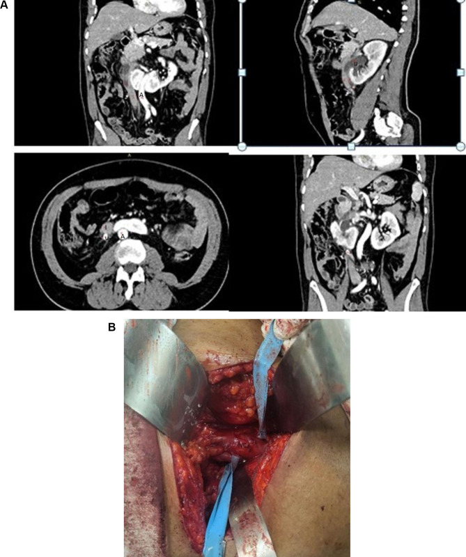
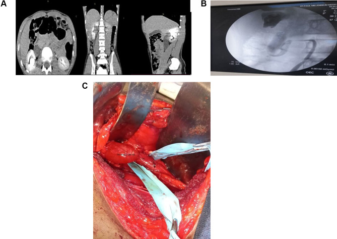
Retrocaval ureter is a rare congenital vascular anomaly described as the passage of the ureter behind the inferior vena cava (IVC) and then turning around the IVC to attain the final lateral position. The condition is usually associated with obstruction in the ipsilateral kidney, causing different degrees of hydronephrosis and complications associated with urinary stasis, such as stone formation. Imaging has a crucial role in the diagnosis and management of retrocaval ureter. CT urography may be the procedure of choice to confirm the diagnosis and avoid retrograde ureteropyelography. Indications for treatment include flank pain, recurrent infection, hydronephrosis, and stone formation due to obstruction. Surgical management is standard and can be done through either an open, laparoscopic, or robotic approach. In this case series, we are going to see two cases of retrocaval ureter in a 56-year-old male and a 14-year-old male child who presented with a right flank of less than a couple of months duration. The first case has an associated horseshoe kidney and a solitary secondary stone. Both cases were surgically managed with open ureteral division, relocation, and ureteroureterostomy. Both have uneventful post-operative follow-ups.
期刊介绍:
Research and Reports in Urology is an international, peer-reviewed, open access, online journal. Publishing original research, reports, editorials, reviews and commentaries on all aspects of adult and pediatric urology in the clinic and laboratory including the following topics: Pathology, pathophysiology of urological disease Investigation and treatment of urological disease Pharmacology of drugs used for the treatment of urological disease Although the main focus of the journal is to publish research and clinical results in humans; preclinical, animal and in vitro studies will be published where they will shed light on disease processes and potential new therapies. Issues of patient safety and quality of care will also be considered.


 分享
分享
 求助内容:
求助内容: 应助结果提醒方式:
 扫码关注我们
