Anirban Sengupta, Feng Wang, Arabinda Mishra, Jamie L Reed, Li Min Chen, John C Gore
{"title":"松鼠猴大脑静息状态功能网络的检测与表征。","authors":"Anirban Sengupta, Feng Wang, Arabinda Mishra, Jamie L Reed, Li Min Chen, John C Gore","doi":"10.1093/texcom/tgad018","DOIUrl":null,"url":null,"abstract":"<p><p>Resting-state fMRI based on analyzing BOLD signals is widely used to derive functional networks in the brain and how they alter during disease or injury conditions. Resting-state networks can also be used to study brain functional connectomes across species, which provides insights into brain evolution. The squirrel monkey (SM) is a non-human primate (NHP) that is widely used as a preclinical model for experimental manipulations to understand the organization and functioning of the brain. We derived resting-state networks from the whole brain of anesthetized SMs using Independent Component Analysis of BOLD acquisitions. We detected 15 anatomically constrained resting-state networks localized in the cortical and subcortical regions as well as in the white-matter. Networks encompassing visual, somatosensory, executive control, sensorimotor, salience and default mode regions, and subcortical networks including the Hippocampus-Amygdala, thalamus, basal-ganglia and brainstem region correspond well with previously detected networks in humans and NHPs. The connectivity pattern between the networks also agrees well with previously reported seed-based resting-state connectivity of SM brain. This study demonstrates that SMs share remarkable homologous network organization with humans and other NHPs, thereby providing strong support for their suitability as a translational animal model for research and additional insight into brain evolution across species.</p>","PeriodicalId":72551,"journal":{"name":"Cerebral cortex communications","volume":"4 3","pages":"tgad018"},"PeriodicalIF":0.0000,"publicationDate":"2023-09-02","publicationTypes":"Journal Article","fieldsOfStudy":null,"isOpenAccess":false,"openAccessPdf":"https://www.ncbi.nlm.nih.gov/pmc/articles/PMC10518810/pdf/","citationCount":"0","resultStr":"{\"title\":\"Detection and characterization of resting state functional networks in squirrel monkey brain.\",\"authors\":\"Anirban Sengupta, Feng Wang, Arabinda Mishra, Jamie L Reed, Li Min Chen, John C Gore\",\"doi\":\"10.1093/texcom/tgad018\",\"DOIUrl\":null,\"url\":null,\"abstract\":\"<p><p>Resting-state fMRI based on analyzing BOLD signals is widely used to derive functional networks in the brain and how they alter during disease or injury conditions. Resting-state networks can also be used to study brain functional connectomes across species, which provides insights into brain evolution. The squirrel monkey (SM) is a non-human primate (NHP) that is widely used as a preclinical model for experimental manipulations to understand the organization and functioning of the brain. We derived resting-state networks from the whole brain of anesthetized SMs using Independent Component Analysis of BOLD acquisitions. We detected 15 anatomically constrained resting-state networks localized in the cortical and subcortical regions as well as in the white-matter. Networks encompassing visual, somatosensory, executive control, sensorimotor, salience and default mode regions, and subcortical networks including the Hippocampus-Amygdala, thalamus, basal-ganglia and brainstem region correspond well with previously detected networks in humans and NHPs. The connectivity pattern between the networks also agrees well with previously reported seed-based resting-state connectivity of SM brain. This study demonstrates that SMs share remarkable homologous network organization with humans and other NHPs, thereby providing strong support for their suitability as a translational animal model for research and additional insight into brain evolution across species.</p>\",\"PeriodicalId\":72551,\"journal\":{\"name\":\"Cerebral cortex communications\",\"volume\":\"4 3\",\"pages\":\"tgad018\"},\"PeriodicalIF\":0.0000,\"publicationDate\":\"2023-09-02\",\"publicationTypes\":\"Journal Article\",\"fieldsOfStudy\":null,\"isOpenAccess\":false,\"openAccessPdf\":\"https://www.ncbi.nlm.nih.gov/pmc/articles/PMC10518810/pdf/\",\"citationCount\":\"0\",\"resultStr\":null,\"platform\":\"Semanticscholar\",\"paperid\":null,\"PeriodicalName\":\"Cerebral cortex communications\",\"FirstCategoryId\":\"1085\",\"ListUrlMain\":\"https://doi.org/10.1093/texcom/tgad018\",\"RegionNum\":0,\"RegionCategory\":null,\"ArticlePicture\":[],\"TitleCN\":null,\"AbstractTextCN\":null,\"PMCID\":null,\"EPubDate\":\"2023/1/1 0:00:00\",\"PubModel\":\"eCollection\",\"JCR\":\"\",\"JCRName\":\"\",\"Score\":null,\"Total\":0}","platform":"Semanticscholar","paperid":null,"PeriodicalName":"Cerebral cortex communications","FirstCategoryId":"1085","ListUrlMain":"https://doi.org/10.1093/texcom/tgad018","RegionNum":0,"RegionCategory":null,"ArticlePicture":[],"TitleCN":null,"AbstractTextCN":null,"PMCID":null,"EPubDate":"2023/1/1 0:00:00","PubModel":"eCollection","JCR":"","JCRName":"","Score":null,"Total":0}
Detection and characterization of resting state functional networks in squirrel monkey brain.
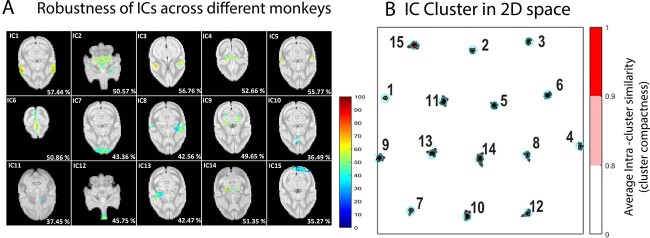
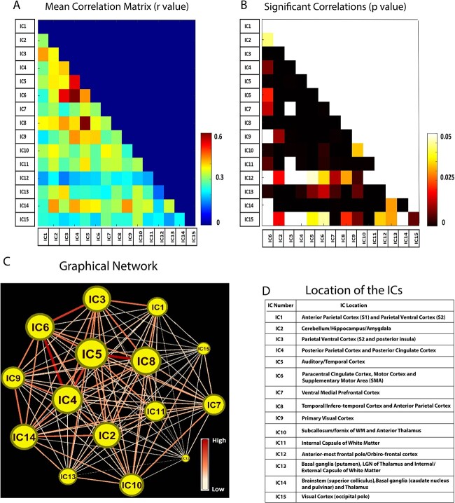
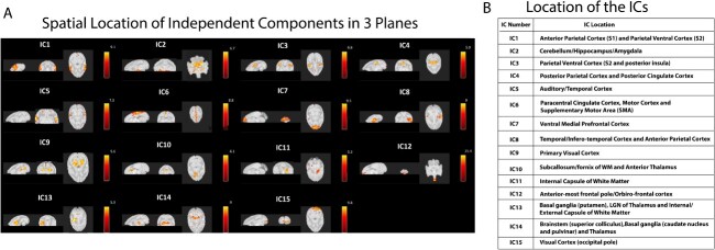
Resting-state fMRI based on analyzing BOLD signals is widely used to derive functional networks in the brain and how they alter during disease or injury conditions. Resting-state networks can also be used to study brain functional connectomes across species, which provides insights into brain evolution. The squirrel monkey (SM) is a non-human primate (NHP) that is widely used as a preclinical model for experimental manipulations to understand the organization and functioning of the brain. We derived resting-state networks from the whole brain of anesthetized SMs using Independent Component Analysis of BOLD acquisitions. We detected 15 anatomically constrained resting-state networks localized in the cortical and subcortical regions as well as in the white-matter. Networks encompassing visual, somatosensory, executive control, sensorimotor, salience and default mode regions, and subcortical networks including the Hippocampus-Amygdala, thalamus, basal-ganglia and brainstem region correspond well with previously detected networks in humans and NHPs. The connectivity pattern between the networks also agrees well with previously reported seed-based resting-state connectivity of SM brain. This study demonstrates that SMs share remarkable homologous network organization with humans and other NHPs, thereby providing strong support for their suitability as a translational animal model for research and additional insight into brain evolution across species.


 分享
分享

 求助内容:
求助内容: 应助结果提醒方式:
 扫码关注我们
