Iuliana Mohorea, Bogdan Socea, Alexandru Constantin Carâp, Dragoş Șerban, Zenaida Ceaușu, Mihail Ceauşu
{"title":"甲状腺肿瘤的形态计量学研究。","authors":"Iuliana Mohorea, Bogdan Socea, Alexandru Constantin Carâp, Dragoş Șerban, Zenaida Ceaușu, Mihail Ceauşu","doi":"10.3892/etm.2023.12196","DOIUrl":null,"url":null,"abstract":"<p><p>Various morphonuclear studies using digital image analysis have been taken into account in order to establish the malignancy of thyroid lesions based on their size and on the chromatographic characteristics of tumor cell nuclei. Nuclear morphometry involves the measurement of nuclear parameters to obtain diagnostically important information in an objective and reproducible manner. The aim of the present study was to evaluate the detailed morphometric analysis of histopathological preparations with lesions of the thyroid gland and to investigate its role in differentiating between benign and malignant thyroid lesions. The present study included 10 benign and 26 malignant thyroid cases with different selected thyroid lesions. Using a microscope connected to a computerized video system, nuclear morphometric parameters including the nuclear area, perimeter, average intensity, red average, width and roundness, were measured and analyzed. The main parameters used in the statistical calculation were significant in distinguishing between benign and malignant thyroid lesions. The association of morphometry in cytological smears for suspected malignant follicular lesions led to increased accuracy in establishing a suspicious malignant diagnosis for follicular lesions. Nuclear morphometry provides an unbiased point of view that increases diagnosis accuracy. Computerized morphometry can positively influence diagnostic accuracy, allowing for a better correlation with clinical and imaging data.</p>","PeriodicalId":94002,"journal":{"name":"Experimental and therapeutic medicine","volume":"26 4","pages":"497"},"PeriodicalIF":2.3000,"publicationDate":"2023-09-06","publicationTypes":"Journal Article","fieldsOfStudy":null,"isOpenAccess":false,"openAccessPdf":"https://ftp.ncbi.nlm.nih.gov/pub/pmc/oa_pdf/ad/5d/etm-26-04-12196.PMC10515107.pdf","citationCount":"0","resultStr":"{\"title\":\"Morphometric study in thyroid tumors.\",\"authors\":\"Iuliana Mohorea, Bogdan Socea, Alexandru Constantin Carâp, Dragoş Șerban, Zenaida Ceaușu, Mihail Ceauşu\",\"doi\":\"10.3892/etm.2023.12196\",\"DOIUrl\":null,\"url\":null,\"abstract\":\"<p><p>Various morphonuclear studies using digital image analysis have been taken into account in order to establish the malignancy of thyroid lesions based on their size and on the chromatographic characteristics of tumor cell nuclei. Nuclear morphometry involves the measurement of nuclear parameters to obtain diagnostically important information in an objective and reproducible manner. The aim of the present study was to evaluate the detailed morphometric analysis of histopathological preparations with lesions of the thyroid gland and to investigate its role in differentiating between benign and malignant thyroid lesions. The present study included 10 benign and 26 malignant thyroid cases with different selected thyroid lesions. Using a microscope connected to a computerized video system, nuclear morphometric parameters including the nuclear area, perimeter, average intensity, red average, width and roundness, were measured and analyzed. The main parameters used in the statistical calculation were significant in distinguishing between benign and malignant thyroid lesions. The association of morphometry in cytological smears for suspected malignant follicular lesions led to increased accuracy in establishing a suspicious malignant diagnosis for follicular lesions. Nuclear morphometry provides an unbiased point of view that increases diagnosis accuracy. Computerized morphometry can positively influence diagnostic accuracy, allowing for a better correlation with clinical and imaging data.</p>\",\"PeriodicalId\":94002,\"journal\":{\"name\":\"Experimental and therapeutic medicine\",\"volume\":\"26 4\",\"pages\":\"497\"},\"PeriodicalIF\":2.3000,\"publicationDate\":\"2023-09-06\",\"publicationTypes\":\"Journal Article\",\"fieldsOfStudy\":null,\"isOpenAccess\":false,\"openAccessPdf\":\"https://ftp.ncbi.nlm.nih.gov/pub/pmc/oa_pdf/ad/5d/etm-26-04-12196.PMC10515107.pdf\",\"citationCount\":\"0\",\"resultStr\":null,\"platform\":\"Semanticscholar\",\"paperid\":null,\"PeriodicalName\":\"Experimental and therapeutic medicine\",\"FirstCategoryId\":\"1085\",\"ListUrlMain\":\"https://doi.org/10.3892/etm.2023.12196\",\"RegionNum\":0,\"RegionCategory\":null,\"ArticlePicture\":[],\"TitleCN\":null,\"AbstractTextCN\":null,\"PMCID\":null,\"EPubDate\":\"2023/10/1 0:00:00\",\"PubModel\":\"eCollection\",\"JCR\":\"\",\"JCRName\":\"\",\"Score\":null,\"Total\":0}","platform":"Semanticscholar","paperid":null,"PeriodicalName":"Experimental and therapeutic medicine","FirstCategoryId":"1085","ListUrlMain":"https://doi.org/10.3892/etm.2023.12196","RegionNum":0,"RegionCategory":null,"ArticlePicture":[],"TitleCN":null,"AbstractTextCN":null,"PMCID":null,"EPubDate":"2023/10/1 0:00:00","PubModel":"eCollection","JCR":"","JCRName":"","Score":null,"Total":0}
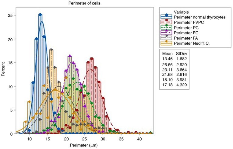
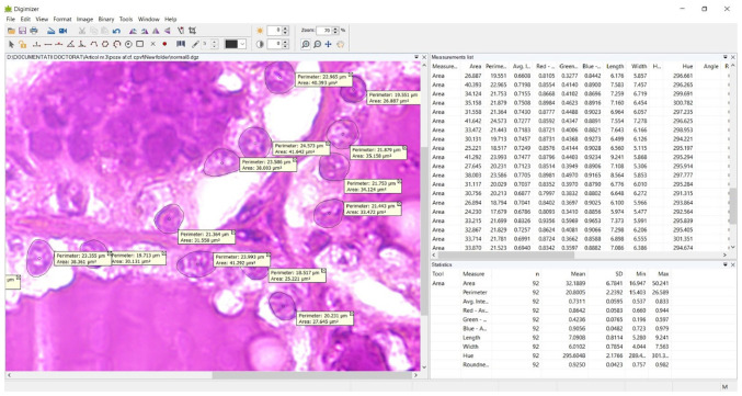
Various morphonuclear studies using digital image analysis have been taken into account in order to establish the malignancy of thyroid lesions based on their size and on the chromatographic characteristics of tumor cell nuclei. Nuclear morphometry involves the measurement of nuclear parameters to obtain diagnostically important information in an objective and reproducible manner. The aim of the present study was to evaluate the detailed morphometric analysis of histopathological preparations with lesions of the thyroid gland and to investigate its role in differentiating between benign and malignant thyroid lesions. The present study included 10 benign and 26 malignant thyroid cases with different selected thyroid lesions. Using a microscope connected to a computerized video system, nuclear morphometric parameters including the nuclear area, perimeter, average intensity, red average, width and roundness, were measured and analyzed. The main parameters used in the statistical calculation were significant in distinguishing between benign and malignant thyroid lesions. The association of morphometry in cytological smears for suspected malignant follicular lesions led to increased accuracy in establishing a suspicious malignant diagnosis for follicular lesions. Nuclear morphometry provides an unbiased point of view that increases diagnosis accuracy. Computerized morphometry can positively influence diagnostic accuracy, allowing for a better correlation with clinical and imaging data.


 分享
分享

 求助内容:
求助内容: 应助结果提醒方式:
 扫码关注我们
