Jiang Cao, Jie Chang, Chaoqin Wu, Sheng Zhang, Binyu Wang, Kaixiang Yang, Xiaojian Cao, Tao Sui
{"title":"硬膜外对侧S1神经根移植治疗痉挛性下肢瘫痪。","authors":"Jiang Cao, Jie Chang, Chaoqin Wu, Sheng Zhang, Binyu Wang, Kaixiang Yang, Xiaojian Cao, Tao Sui","doi":"10.7555/JBR.37.20230068","DOIUrl":null,"url":null,"abstract":"<p><p>The current study aims to ascertain the anatomical feasibility of transferring the contralateral S1 ventral root (VR) to the ipsilateral L5 VR for treating unilateral spastic lower limb paralysis. Six formalin-fixed (three males and three females) cadavers were used. The VR of the contralateral S1 was transferred to the VR of the ipsilateral L5. The sural nerve was selected as a bridge between the donor and recipient nerve. The number of axons, the cross-sectional areas and the pertinent distances between the donor and recipient nerves were measured. The extradural S1 VR and L5 VR could be separated based on anatomical markers of the dorsal root ganglion. The gross distance between the S1 nerve root and L5 nerve root was 31.31 (± 3.23) mm in the six cadavers, while that on the diffusion tensor imaging was 47.51 (± 3.23) mm in 60 patients without spinal diseases, and both distances were seperately greater than that between the outlet of S1 from the spinal cord and the ganglion. The numbers of axons in the S1 VRs and L5 VRs were 13414.20 (± 2890.30) and 10613.20 (± 2135.58), respectively. The cross-sectional areas of the S1 VR and L5 VR were 1.68 (± 0.26) mm <sup>2</sup> and 1.08 (± 0.26) mm <sup>2</sup>, respectively. In conclusion, transfer of the contralateral S1 VR to the ipsilateral L5 VR may be an anatomically feasible treatment option for unilateral spastic lower limb paralysis.</p>","PeriodicalId":15061,"journal":{"name":"Journal of Biomedical Research","volume":" ","pages":"394-400"},"PeriodicalIF":2.2000,"publicationDate":"2023-09-28","publicationTypes":"Journal Article","fieldsOfStudy":null,"isOpenAccess":false,"openAccessPdf":"https://www.ncbi.nlm.nih.gov/pmc/articles/PMC10541774/pdf/","citationCount":"0","resultStr":"{\"title\":\"Extradural contralateral S1 nerve root transfer for spastic lower limb paralysis.\",\"authors\":\"Jiang Cao, Jie Chang, Chaoqin Wu, Sheng Zhang, Binyu Wang, Kaixiang Yang, Xiaojian Cao, Tao Sui\",\"doi\":\"10.7555/JBR.37.20230068\",\"DOIUrl\":null,\"url\":null,\"abstract\":\"<p><p>The current study aims to ascertain the anatomical feasibility of transferring the contralateral S1 ventral root (VR) to the ipsilateral L5 VR for treating unilateral spastic lower limb paralysis. Six formalin-fixed (three males and three females) cadavers were used. The VR of the contralateral S1 was transferred to the VR of the ipsilateral L5. The sural nerve was selected as a bridge between the donor and recipient nerve. The number of axons, the cross-sectional areas and the pertinent distances between the donor and recipient nerves were measured. The extradural S1 VR and L5 VR could be separated based on anatomical markers of the dorsal root ganglion. The gross distance between the S1 nerve root and L5 nerve root was 31.31 (± 3.23) mm in the six cadavers, while that on the diffusion tensor imaging was 47.51 (± 3.23) mm in 60 patients without spinal diseases, and both distances were seperately greater than that between the outlet of S1 from the spinal cord and the ganglion. The numbers of axons in the S1 VRs and L5 VRs were 13414.20 (± 2890.30) and 10613.20 (± 2135.58), respectively. The cross-sectional areas of the S1 VR and L5 VR were 1.68 (± 0.26) mm <sup>2</sup> and 1.08 (± 0.26) mm <sup>2</sup>, respectively. In conclusion, transfer of the contralateral S1 VR to the ipsilateral L5 VR may be an anatomically feasible treatment option for unilateral spastic lower limb paralysis.</p>\",\"PeriodicalId\":15061,\"journal\":{\"name\":\"Journal of Biomedical Research\",\"volume\":\" \",\"pages\":\"394-400\"},\"PeriodicalIF\":2.2000,\"publicationDate\":\"2023-09-28\",\"publicationTypes\":\"Journal Article\",\"fieldsOfStudy\":null,\"isOpenAccess\":false,\"openAccessPdf\":\"https://www.ncbi.nlm.nih.gov/pmc/articles/PMC10541774/pdf/\",\"citationCount\":\"0\",\"resultStr\":null,\"platform\":\"Semanticscholar\",\"paperid\":null,\"PeriodicalName\":\"Journal of Biomedical Research\",\"FirstCategoryId\":\"1087\",\"ListUrlMain\":\"https://doi.org/10.7555/JBR.37.20230068\",\"RegionNum\":4,\"RegionCategory\":\"医学\",\"ArticlePicture\":[],\"TitleCN\":null,\"AbstractTextCN\":null,\"PMCID\":null,\"EPubDate\":\"\",\"PubModel\":\"\",\"JCR\":\"Q3\",\"JCRName\":\"MEDICINE, RESEARCH & EXPERIMENTAL\",\"Score\":null,\"Total\":0}","platform":"Semanticscholar","paperid":null,"PeriodicalName":"Journal of Biomedical Research","FirstCategoryId":"1087","ListUrlMain":"https://doi.org/10.7555/JBR.37.20230068","RegionNum":4,"RegionCategory":"医学","ArticlePicture":[],"TitleCN":null,"AbstractTextCN":null,"PMCID":null,"EPubDate":"","PubModel":"","JCR":"Q3","JCRName":"MEDICINE, RESEARCH & EXPERIMENTAL","Score":null,"Total":0}
Extradural contralateral S1 nerve root transfer for spastic lower limb paralysis.
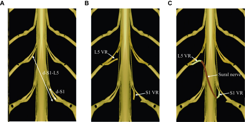
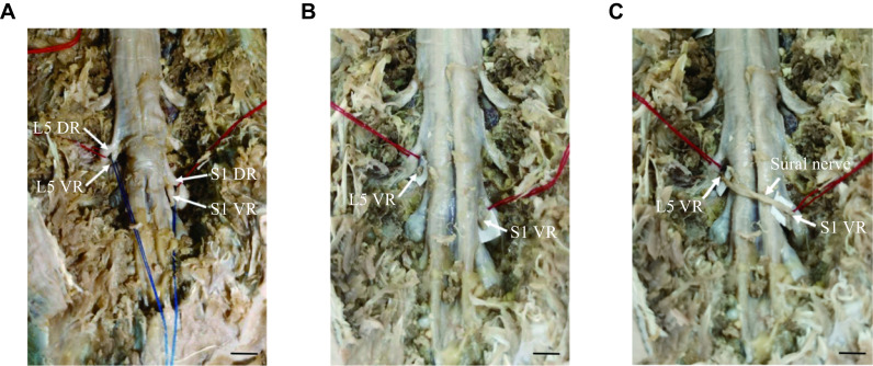
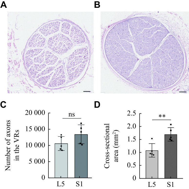
The current study aims to ascertain the anatomical feasibility of transferring the contralateral S1 ventral root (VR) to the ipsilateral L5 VR for treating unilateral spastic lower limb paralysis. Six formalin-fixed (three males and three females) cadavers were used. The VR of the contralateral S1 was transferred to the VR of the ipsilateral L5. The sural nerve was selected as a bridge between the donor and recipient nerve. The number of axons, the cross-sectional areas and the pertinent distances between the donor and recipient nerves were measured. The extradural S1 VR and L5 VR could be separated based on anatomical markers of the dorsal root ganglion. The gross distance between the S1 nerve root and L5 nerve root was 31.31 (± 3.23) mm in the six cadavers, while that on the diffusion tensor imaging was 47.51 (± 3.23) mm in 60 patients without spinal diseases, and both distances were seperately greater than that between the outlet of S1 from the spinal cord and the ganglion. The numbers of axons in the S1 VRs and L5 VRs were 13414.20 (± 2890.30) and 10613.20 (± 2135.58), respectively. The cross-sectional areas of the S1 VR and L5 VR were 1.68 (± 0.26) mm 2 and 1.08 (± 0.26) mm 2, respectively. In conclusion, transfer of the contralateral S1 VR to the ipsilateral L5 VR may be an anatomically feasible treatment option for unilateral spastic lower limb paralysis.


 分享
分享
 求助内容:
求助内容: 应助结果提醒方式:
 扫码关注我们
