Alexandra Brown, Brooklyn Morris, John Karanja Kamau, Abdullah A Alshudukhi, Abdulrahman Jama, Hongmei Ren
{"title":"使用细胞剖面仪监测肌肉营养不良疾病进展的自动图像分析管道开发。","authors":"Alexandra Brown, Brooklyn Morris, John Karanja Kamau, Abdullah A Alshudukhi, Abdulrahman Jama, Hongmei Ren","doi":"10.26502/ami.936500115","DOIUrl":null,"url":null,"abstract":"<p><p>Muscular dystrophies are inherited disorders that are characterized by progressive muscle degeneration. These disorders are caused by mutations in the genes encoding structural elements within the muscle, which leads to increased vulnerability to mechanical stress and sarcolemma damage. Although myofibers have the capacity to regenerate, the newly formed myofibers still harbor genetic mutation, which induces continuous cycles of muscle fiber death and regeneration. This repeated cycling is accompanied by an inflammatory response which eventually provokes excessive fibrotic deposition. The histopathological changes in skeletal muscle tissue are central to the disease pathogenesis. Analysis of muscle histopathology is the gold standard for monitoring muscle health and disease progression. However, manual, or semi-manual quantification methods, are not only immensely tedious but can be subjective. Here, we present four image analysis pipelines built in CellProfiler which enable users without a background in computer vision or programming to quantitatively analyze biological images. These image analysis pipelines are designed to quantify skeletal muscle histopathological staining for membrane damage, the abundance and size distribution of regenerating muscle fibers, inflammation via quantification of CD68+ M1 macrophages, and collagen deposition. Additionally, we discuss methods to address common errors associated with the quantification of microscopy images. These automated tools can not only improve workflow efficiency but can provide a better understanding of the histopathological progression of muscular dystrophy.</p>","PeriodicalId":72285,"journal":{"name":"Archives of microbiology & immunology","volume":"7 3","pages":"178-187"},"PeriodicalIF":0.0000,"publicationDate":"2023-01-01","publicationTypes":"Journal Article","fieldsOfStudy":null,"isOpenAccess":false,"openAccessPdf":"https://www.ncbi.nlm.nih.gov/pmc/articles/PMC10552673/pdf/","citationCount":"0","resultStr":"{\"title\":\"Automated Image Analysis Pipeline Development to Monitor Disease Progression in Muscular Dystrophy Using Cell Profiler.\",\"authors\":\"Alexandra Brown, Brooklyn Morris, John Karanja Kamau, Abdullah A Alshudukhi, Abdulrahman Jama, Hongmei Ren\",\"doi\":\"10.26502/ami.936500115\",\"DOIUrl\":null,\"url\":null,\"abstract\":\"<p><p>Muscular dystrophies are inherited disorders that are characterized by progressive muscle degeneration. These disorders are caused by mutations in the genes encoding structural elements within the muscle, which leads to increased vulnerability to mechanical stress and sarcolemma damage. Although myofibers have the capacity to regenerate, the newly formed myofibers still harbor genetic mutation, which induces continuous cycles of muscle fiber death and regeneration. This repeated cycling is accompanied by an inflammatory response which eventually provokes excessive fibrotic deposition. The histopathological changes in skeletal muscle tissue are central to the disease pathogenesis. Analysis of muscle histopathology is the gold standard for monitoring muscle health and disease progression. However, manual, or semi-manual quantification methods, are not only immensely tedious but can be subjective. Here, we present four image analysis pipelines built in CellProfiler which enable users without a background in computer vision or programming to quantitatively analyze biological images. These image analysis pipelines are designed to quantify skeletal muscle histopathological staining for membrane damage, the abundance and size distribution of regenerating muscle fibers, inflammation via quantification of CD68+ M1 macrophages, and collagen deposition. Additionally, we discuss methods to address common errors associated with the quantification of microscopy images. These automated tools can not only improve workflow efficiency but can provide a better understanding of the histopathological progression of muscular dystrophy.</p>\",\"PeriodicalId\":72285,\"journal\":{\"name\":\"Archives of microbiology & immunology\",\"volume\":\"7 3\",\"pages\":\"178-187\"},\"PeriodicalIF\":0.0000,\"publicationDate\":\"2023-01-01\",\"publicationTypes\":\"Journal Article\",\"fieldsOfStudy\":null,\"isOpenAccess\":false,\"openAccessPdf\":\"https://www.ncbi.nlm.nih.gov/pmc/articles/PMC10552673/pdf/\",\"citationCount\":\"0\",\"resultStr\":null,\"platform\":\"Semanticscholar\",\"paperid\":null,\"PeriodicalName\":\"Archives of microbiology & immunology\",\"FirstCategoryId\":\"1085\",\"ListUrlMain\":\"https://doi.org/10.26502/ami.936500115\",\"RegionNum\":0,\"RegionCategory\":null,\"ArticlePicture\":[],\"TitleCN\":null,\"AbstractTextCN\":null,\"PMCID\":null,\"EPubDate\":\"2023/9/1 0:00:00\",\"PubModel\":\"Epub\",\"JCR\":\"\",\"JCRName\":\"\",\"Score\":null,\"Total\":0}","platform":"Semanticscholar","paperid":null,"PeriodicalName":"Archives of microbiology & immunology","FirstCategoryId":"1085","ListUrlMain":"https://doi.org/10.26502/ami.936500115","RegionNum":0,"RegionCategory":null,"ArticlePicture":[],"TitleCN":null,"AbstractTextCN":null,"PMCID":null,"EPubDate":"2023/9/1 0:00:00","PubModel":"Epub","JCR":"","JCRName":"","Score":null,"Total":0}
Automated Image Analysis Pipeline Development to Monitor Disease Progression in Muscular Dystrophy Using Cell Profiler.
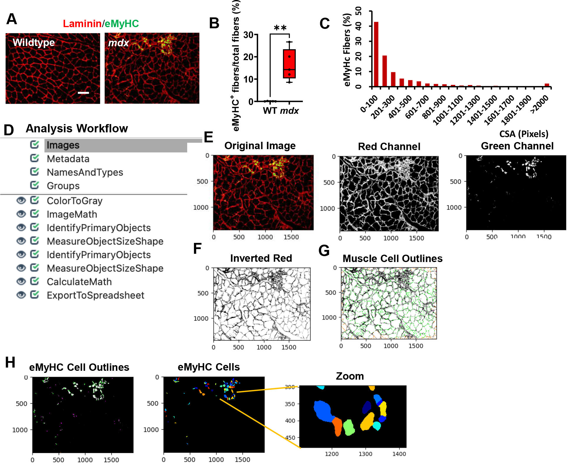
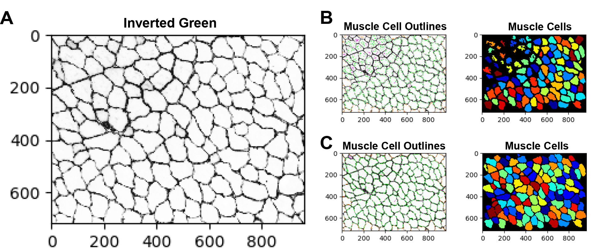
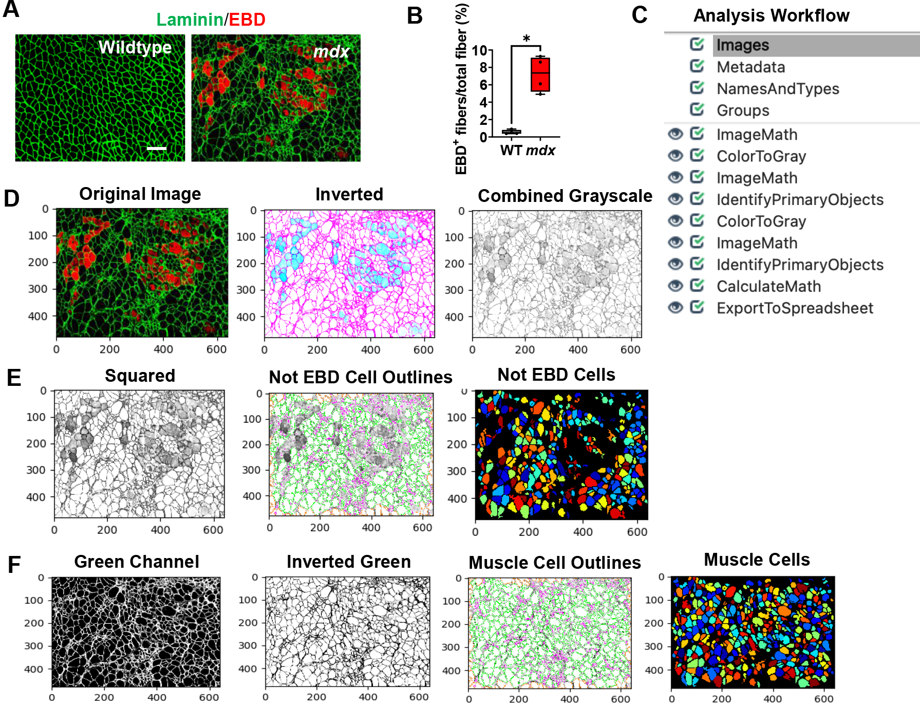
Muscular dystrophies are inherited disorders that are characterized by progressive muscle degeneration. These disorders are caused by mutations in the genes encoding structural elements within the muscle, which leads to increased vulnerability to mechanical stress and sarcolemma damage. Although myofibers have the capacity to regenerate, the newly formed myofibers still harbor genetic mutation, which induces continuous cycles of muscle fiber death and regeneration. This repeated cycling is accompanied by an inflammatory response which eventually provokes excessive fibrotic deposition. The histopathological changes in skeletal muscle tissue are central to the disease pathogenesis. Analysis of muscle histopathology is the gold standard for monitoring muscle health and disease progression. However, manual, or semi-manual quantification methods, are not only immensely tedious but can be subjective. Here, we present four image analysis pipelines built in CellProfiler which enable users without a background in computer vision or programming to quantitatively analyze biological images. These image analysis pipelines are designed to quantify skeletal muscle histopathological staining for membrane damage, the abundance and size distribution of regenerating muscle fibers, inflammation via quantification of CD68+ M1 macrophages, and collagen deposition. Additionally, we discuss methods to address common errors associated with the quantification of microscopy images. These automated tools can not only improve workflow efficiency but can provide a better understanding of the histopathological progression of muscular dystrophy.


 分享
分享

 求助内容:
求助内容: 应助结果提醒方式:
 扫码关注我们
