Yuanyuan Sun, Chunfeng Xu, Mingjie Wang, Lingfei Wei, Herman Pieterse, Yiqun Wu, Yuelian Liu
{"title":"rhBMP-2结合的仿生磷酸钙材料在临床牙槽窝保存中诱导骨形成的放射学和组织学评估。","authors":"Yuanyuan Sun, Chunfeng Xu, Mingjie Wang, Lingfei Wei, Herman Pieterse, Yiqun Wu, Yuelian Liu","doi":"10.1186/s40729-023-00491-1","DOIUrl":null,"url":null,"abstract":"<p><strong>Purpose: </strong>We assessed the efficiency of low-dose recombinant human bone morphogenetic protein-2 (rhBMP-2) incorporated biomimetic calcium phosphate on β-tricalcium phosphate (β-TCP) (rhBMP-2/BioCaP/β-TCP) on bone formation in a model of socket preservation using cone beam computed tomography (CBCT) scanning and histological examination.</p><p><strong>Methods: </strong>Forty patients undergoing minimally invasive single-root tooth extraction for dental implantation were randomized to three groups according to the material used for socket preservation: filling with rhBMP-2/BioCaP/β-TCP, β-TCP, or natural healing (kept unfilled) (controls). The alveolar sockets (including the control group) were covered by two-layer collagen membranes and sutured. Two CBCT scans were taken, one immediately after socket preservation procedure (baseline) and another 6 weeks later. Gray values (GVs) obtained from CBCT were recorded. During insertion of the dental implant, biopsies were taken and analyzed histologically for new bone formation, residual material, and unmineralized bone tissue at the core of the biopsy.</p><p><strong>Results: </strong>The mean (± standard deviation) changes of GVs of the CBCT scans at the central area of filled materials were as follows: 373.19 ± 157.16 in the rhBMP-2/BioCaP/β-TCP group, 112.26 ± 197.25 in the β-TCP group, and -257 ± 273.51 in the control group. The decrease of GVs in the rhBMP-2/BioCaP/β-TCP group as compared with the β-TCP group was statistically significant (P < 0.001). Differences in new bone formation (P = 0.006) were also found: 21,18% ± 7.62% in the rhBMP-2/BioCaP/β-TCP group, 13.44% ± 6.03% in the β-TCP group, and 9.49% ± 0.08% in controls. The residual material was10.04% ± 4.57% in the rhBMP-2/BioCaP/β-TCP group vs. 20.60% ± 9.54%) in the β-TCP group (P < 0.001). Differences in unmineralized bone tissue (P < 0.001) were also found (68.78% ± 7.67%, 65.96% ± 12.64%, and 90.38% ± 7.5% in the rhBMP-2/BioCaP/β-TC, β-TCP, and control groups, respectively).</p><p><strong>Conclusions: </strong>This study shows that rhBMP-2/BioCaP/β-TCP is a promising bone substitute with fast degradation and potent pro-osteogenic capacity that can be useful for socket preservation in implant dentistry.</p><p><strong>Trial registration: </strong>ChiCTR, ChiCTR2000035263. Registered 5 August 2020, https://www.chictr.org.cn/ChiCTR2000035263 .</p>","PeriodicalId":14076,"journal":{"name":"International Journal of Implant Dentistry","volume":"9 1","pages":"37"},"PeriodicalIF":4.0000,"publicationDate":"2023-10-16","publicationTypes":"Journal Article","fieldsOfStudy":null,"isOpenAccess":false,"openAccessPdf":"https://www.ncbi.nlm.nih.gov/pmc/articles/PMC10579201/pdf/","citationCount":"0","resultStr":"{\"title\":\"Radiographic and histological evaluation of bone formation induced by rhBMP-2-incorporated biomimetic calcium phosphate material in clinical alveolar sockets preservation.\",\"authors\":\"Yuanyuan Sun, Chunfeng Xu, Mingjie Wang, Lingfei Wei, Herman Pieterse, Yiqun Wu, Yuelian Liu\",\"doi\":\"10.1186/s40729-023-00491-1\",\"DOIUrl\":null,\"url\":null,\"abstract\":\"<p><strong>Purpose: </strong>We assessed the efficiency of low-dose recombinant human bone morphogenetic protein-2 (rhBMP-2) incorporated biomimetic calcium phosphate on β-tricalcium phosphate (β-TCP) (rhBMP-2/BioCaP/β-TCP) on bone formation in a model of socket preservation using cone beam computed tomography (CBCT) scanning and histological examination.</p><p><strong>Methods: </strong>Forty patients undergoing minimally invasive single-root tooth extraction for dental implantation were randomized to three groups according to the material used for socket preservation: filling with rhBMP-2/BioCaP/β-TCP, β-TCP, or natural healing (kept unfilled) (controls). The alveolar sockets (including the control group) were covered by two-layer collagen membranes and sutured. Two CBCT scans were taken, one immediately after socket preservation procedure (baseline) and another 6 weeks later. Gray values (GVs) obtained from CBCT were recorded. During insertion of the dental implant, biopsies were taken and analyzed histologically for new bone formation, residual material, and unmineralized bone tissue at the core of the biopsy.</p><p><strong>Results: </strong>The mean (± standard deviation) changes of GVs of the CBCT scans at the central area of filled materials were as follows: 373.19 ± 157.16 in the rhBMP-2/BioCaP/β-TCP group, 112.26 ± 197.25 in the β-TCP group, and -257 ± 273.51 in the control group. The decrease of GVs in the rhBMP-2/BioCaP/β-TCP group as compared with the β-TCP group was statistically significant (P < 0.001). Differences in new bone formation (P = 0.006) were also found: 21,18% ± 7.62% in the rhBMP-2/BioCaP/β-TCP group, 13.44% ± 6.03% in the β-TCP group, and 9.49% ± 0.08% in controls. The residual material was10.04% ± 4.57% in the rhBMP-2/BioCaP/β-TCP group vs. 20.60% ± 9.54%) in the β-TCP group (P < 0.001). Differences in unmineralized bone tissue (P < 0.001) were also found (68.78% ± 7.67%, 65.96% ± 12.64%, and 90.38% ± 7.5% in the rhBMP-2/BioCaP/β-TC, β-TCP, and control groups, respectively).</p><p><strong>Conclusions: </strong>This study shows that rhBMP-2/BioCaP/β-TCP is a promising bone substitute with fast degradation and potent pro-osteogenic capacity that can be useful for socket preservation in implant dentistry.</p><p><strong>Trial registration: </strong>ChiCTR, ChiCTR2000035263. Registered 5 August 2020, https://www.chictr.org.cn/ChiCTR2000035263 .</p>\",\"PeriodicalId\":14076,\"journal\":{\"name\":\"International Journal of Implant Dentistry\",\"volume\":\"9 1\",\"pages\":\"37\"},\"PeriodicalIF\":4.0000,\"publicationDate\":\"2023-10-16\",\"publicationTypes\":\"Journal Article\",\"fieldsOfStudy\":null,\"isOpenAccess\":false,\"openAccessPdf\":\"https://www.ncbi.nlm.nih.gov/pmc/articles/PMC10579201/pdf/\",\"citationCount\":\"0\",\"resultStr\":null,\"platform\":\"Semanticscholar\",\"paperid\":null,\"PeriodicalName\":\"International Journal of Implant Dentistry\",\"FirstCategoryId\":\"3\",\"ListUrlMain\":\"https://doi.org/10.1186/s40729-023-00491-1\",\"RegionNum\":3,\"RegionCategory\":\"医学\",\"ArticlePicture\":[],\"TitleCN\":null,\"AbstractTextCN\":null,\"PMCID\":null,\"EPubDate\":\"\",\"PubModel\":\"\",\"JCR\":\"Q1\",\"JCRName\":\"DENTISTRY, ORAL SURGERY & MEDICINE\",\"Score\":null,\"Total\":0}","platform":"Semanticscholar","paperid":null,"PeriodicalName":"International Journal of Implant Dentistry","FirstCategoryId":"3","ListUrlMain":"https://doi.org/10.1186/s40729-023-00491-1","RegionNum":3,"RegionCategory":"医学","ArticlePicture":[],"TitleCN":null,"AbstractTextCN":null,"PMCID":null,"EPubDate":"","PubModel":"","JCR":"Q1","JCRName":"DENTISTRY, ORAL SURGERY & MEDICINE","Score":null,"Total":0}
Radiographic and histological evaluation of bone formation induced by rhBMP-2-incorporated biomimetic calcium phosphate material in clinical alveolar sockets preservation.
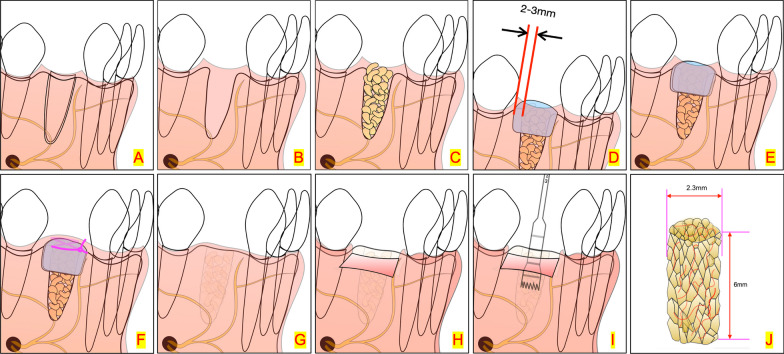
Purpose: We assessed the efficiency of low-dose recombinant human bone morphogenetic protein-2 (rhBMP-2) incorporated biomimetic calcium phosphate on β-tricalcium phosphate (β-TCP) (rhBMP-2/BioCaP/β-TCP) on bone formation in a model of socket preservation using cone beam computed tomography (CBCT) scanning and histological examination.
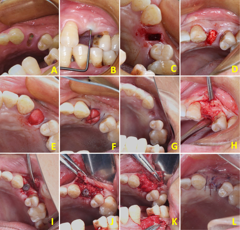
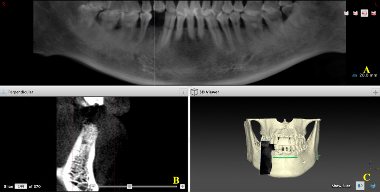
Methods: Forty patients undergoing minimally invasive single-root tooth extraction for dental implantation were randomized to three groups according to the material used for socket preservation: filling with rhBMP-2/BioCaP/β-TCP, β-TCP, or natural healing (kept unfilled) (controls). The alveolar sockets (including the control group) were covered by two-layer collagen membranes and sutured. Two CBCT scans were taken, one immediately after socket preservation procedure (baseline) and another 6 weeks later. Gray values (GVs) obtained from CBCT were recorded. During insertion of the dental implant, biopsies were taken and analyzed histologically for new bone formation, residual material, and unmineralized bone tissue at the core of the biopsy.
Results: The mean (± standard deviation) changes of GVs of the CBCT scans at the central area of filled materials were as follows: 373.19 ± 157.16 in the rhBMP-2/BioCaP/β-TCP group, 112.26 ± 197.25 in the β-TCP group, and -257 ± 273.51 in the control group. The decrease of GVs in the rhBMP-2/BioCaP/β-TCP group as compared with the β-TCP group was statistically significant (P < 0.001). Differences in new bone formation (P = 0.006) were also found: 21,18% ± 7.62% in the rhBMP-2/BioCaP/β-TCP group, 13.44% ± 6.03% in the β-TCP group, and 9.49% ± 0.08% in controls. The residual material was10.04% ± 4.57% in the rhBMP-2/BioCaP/β-TCP group vs. 20.60% ± 9.54%) in the β-TCP group (P < 0.001). Differences in unmineralized bone tissue (P < 0.001) were also found (68.78% ± 7.67%, 65.96% ± 12.64%, and 90.38% ± 7.5% in the rhBMP-2/BioCaP/β-TC, β-TCP, and control groups, respectively).
Conclusions: This study shows that rhBMP-2/BioCaP/β-TCP is a promising bone substitute with fast degradation and potent pro-osteogenic capacity that can be useful for socket preservation in implant dentistry.
Trial registration: ChiCTR, ChiCTR2000035263. Registered 5 August 2020, https://www.chictr.org.cn/ChiCTR2000035263 .
期刊介绍:
The International Journal of Implant Dentistry is a peer-reviewed open access journal published under the SpringerOpen brand. The journal is dedicated to promoting the exchange and discussion of all research areas relevant to implant dentistry in the form of systematic literature or invited reviews, prospective and retrospective clinical studies, clinical case reports, basic laboratory and animal research, and articles on material research and engineering.


 分享
分享
 求助内容:
求助内容: 应助结果提醒方式:
 扫码关注我们
