{"title":"智能手机数码照片上拇外翻角度测量的有效性和可靠性。","authors":"Albert Cakar, Ozkan Kose, Firat Dogruoz, Huseyin Selcuk, Tolga Kirtis, Omer Faruk Egerci","doi":"10.1186/s13047-023-00670-8","DOIUrl":null,"url":null,"abstract":"<p><strong>Background: </strong>This prospective study aimed to test the reliability and validity of hallux valgus angle (HVA) measurement on smartphone digital photographs compared with the standard radiographic evaluation.</p><p><strong>Methods: </strong>Twenty Seven female patients (45 feet) with forefoot deformity were evaluated with weight-bearing anteroposterior foot radiographs and smartphone photographs. Radiographic hallux valgus angle (rHVA) was measured on digital radiographs. Two different photographic HVA measurement methods were used. In the first, the longitudinal axes of the first metatarsal and proximal phalanx were determined, and the angle between these axes was measured (pHVA), similar to the radiographic method. In the other method, the angle of the margo medialis pedis was measured on the photograph (pMMP). Two independent observers performed all measurements twice on two different occasions. Reliability analysis was performed using the interclass correlation coefficient. Agreement between the measurements was tested using Bland-Altman analysis.</p><p><strong>Results: </strong>The repeated rHVA, pHVA and pMMP measurements showed excellent intra and inter-observer reliability, with ICC values above 0.900. The mean rHVA, pHVA, and pMMP were statistically similar (p:0.929, 27.03°±8.7°, 27.11°±8.8° and 26.5°±9.0° respectively). The mean difference between the rHVA and pHVA was - 0.07°±5.1° (range, --9.67 to 9.56°), and the mean difference between the rHVA and pMMP was 0.53°±4.4° (range, -9.76° to 8.22°). There was a strong positive correlation between both photographic methods and radiographic measurements (rho = 0.809, p = 0.001 and rho = 0.872, p = 0.001). In the Bland Altman plot, the upper and lower LOAs (95%CI) ranged from - 10.11° to 9.93° for rHVA and pHVA, and from - 8.26° to 9.33° for rHVA and pMMP. Linear regression analysis showed a proportional bias for pHVA but not for the pMMP (p:0.010 versus p:0.633, respectively). The range of the mean difference (prediction interval) between the pMMP and rHVA was 17.59° and 20° for pHVA and rHVA. Simple linear regression showed that the rHVA was predicted by the following equation: rHVA = 4.73 + 0.84 × pMMP (r2 = 0.761, p < 0.001).</p><p><strong>Conclusions: </strong>Although measuring HVA through smartphone photographs is reliable, it is not a valid prediction method.</p><p><strong>Level of evidence: </strong>Level II, diagnostic assessment.</p>","PeriodicalId":49164,"journal":{"name":"Journal of Foot and Ankle Research","volume":"16 1","pages":"70"},"PeriodicalIF":2.2000,"publicationDate":"2023-10-16","publicationTypes":"Journal Article","fieldsOfStudy":null,"isOpenAccess":false,"openAccessPdf":"https://www.ncbi.nlm.nih.gov/pmc/articles/PMC10577965/pdf/","citationCount":"0","resultStr":"{\"title\":\"Validity and reliability of hallux valgus angle measurement on smartphone digital photographs.\",\"authors\":\"Albert Cakar, Ozkan Kose, Firat Dogruoz, Huseyin Selcuk, Tolga Kirtis, Omer Faruk Egerci\",\"doi\":\"10.1186/s13047-023-00670-8\",\"DOIUrl\":null,\"url\":null,\"abstract\":\"<p><strong>Background: </strong>This prospective study aimed to test the reliability and validity of hallux valgus angle (HVA) measurement on smartphone digital photographs compared with the standard radiographic evaluation.</p><p><strong>Methods: </strong>Twenty Seven female patients (45 feet) with forefoot deformity were evaluated with weight-bearing anteroposterior foot radiographs and smartphone photographs. Radiographic hallux valgus angle (rHVA) was measured on digital radiographs. Two different photographic HVA measurement methods were used. In the first, the longitudinal axes of the first metatarsal and proximal phalanx were determined, and the angle between these axes was measured (pHVA), similar to the radiographic method. In the other method, the angle of the margo medialis pedis was measured on the photograph (pMMP). Two independent observers performed all measurements twice on two different occasions. Reliability analysis was performed using the interclass correlation coefficient. Agreement between the measurements was tested using Bland-Altman analysis.</p><p><strong>Results: </strong>The repeated rHVA, pHVA and pMMP measurements showed excellent intra and inter-observer reliability, with ICC values above 0.900. The mean rHVA, pHVA, and pMMP were statistically similar (p:0.929, 27.03°±8.7°, 27.11°±8.8° and 26.5°±9.0° respectively). The mean difference between the rHVA and pHVA was - 0.07°±5.1° (range, --9.67 to 9.56°), and the mean difference between the rHVA and pMMP was 0.53°±4.4° (range, -9.76° to 8.22°). There was a strong positive correlation between both photographic methods and radiographic measurements (rho = 0.809, p = 0.001 and rho = 0.872, p = 0.001). In the Bland Altman plot, the upper and lower LOAs (95%CI) ranged from - 10.11° to 9.93° for rHVA and pHVA, and from - 8.26° to 9.33° for rHVA and pMMP. Linear regression analysis showed a proportional bias for pHVA but not for the pMMP (p:0.010 versus p:0.633, respectively). The range of the mean difference (prediction interval) between the pMMP and rHVA was 17.59° and 20° for pHVA and rHVA. Simple linear regression showed that the rHVA was predicted by the following equation: rHVA = 4.73 + 0.84 × pMMP (r2 = 0.761, p < 0.001).</p><p><strong>Conclusions: </strong>Although measuring HVA through smartphone photographs is reliable, it is not a valid prediction method.</p><p><strong>Level of evidence: </strong>Level II, diagnostic assessment.</p>\",\"PeriodicalId\":49164,\"journal\":{\"name\":\"Journal of Foot and Ankle Research\",\"volume\":\"16 1\",\"pages\":\"70\"},\"PeriodicalIF\":2.2000,\"publicationDate\":\"2023-10-16\",\"publicationTypes\":\"Journal Article\",\"fieldsOfStudy\":null,\"isOpenAccess\":false,\"openAccessPdf\":\"https://www.ncbi.nlm.nih.gov/pmc/articles/PMC10577965/pdf/\",\"citationCount\":\"0\",\"resultStr\":null,\"platform\":\"Semanticscholar\",\"paperid\":null,\"PeriodicalName\":\"Journal of Foot and Ankle Research\",\"FirstCategoryId\":\"3\",\"ListUrlMain\":\"https://doi.org/10.1186/s13047-023-00670-8\",\"RegionNum\":3,\"RegionCategory\":\"医学\",\"ArticlePicture\":[],\"TitleCN\":null,\"AbstractTextCN\":null,\"PMCID\":null,\"EPubDate\":\"\",\"PubModel\":\"\",\"JCR\":\"Q1\",\"JCRName\":\"ORTHOPEDICS\",\"Score\":null,\"Total\":0}","platform":"Semanticscholar","paperid":null,"PeriodicalName":"Journal of Foot and Ankle Research","FirstCategoryId":"3","ListUrlMain":"https://doi.org/10.1186/s13047-023-00670-8","RegionNum":3,"RegionCategory":"医学","ArticlePicture":[],"TitleCN":null,"AbstractTextCN":null,"PMCID":null,"EPubDate":"","PubModel":"","JCR":"Q1","JCRName":"ORTHOPEDICS","Score":null,"Total":0}
Validity and reliability of hallux valgus angle measurement on smartphone digital photographs.
Background: This prospective study aimed to test the reliability and validity of hallux valgus angle (HVA) measurement on smartphone digital photographs compared with the standard radiographic evaluation.
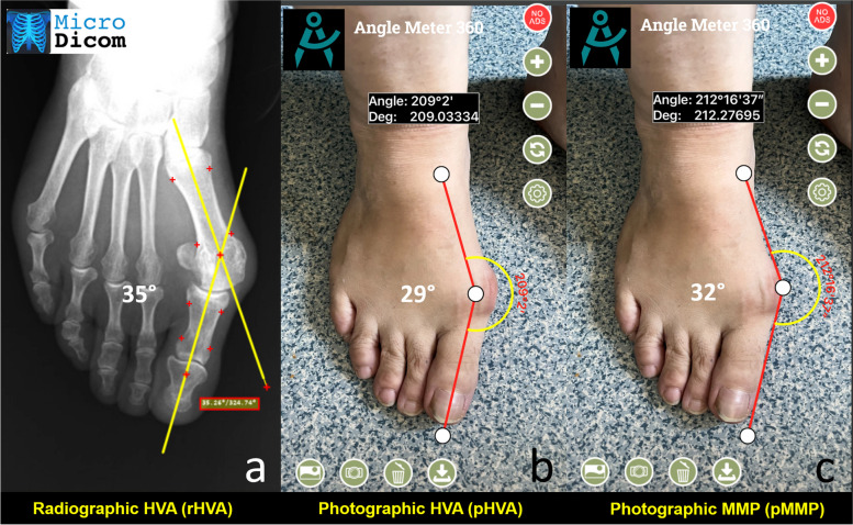
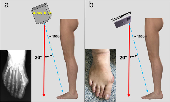
Methods: Twenty Seven female patients (45 feet) with forefoot deformity were evaluated with weight-bearing anteroposterior foot radiographs and smartphone photographs. Radiographic hallux valgus angle (rHVA) was measured on digital radiographs. Two different photographic HVA measurement methods were used. In the first, the longitudinal axes of the first metatarsal and proximal phalanx were determined, and the angle between these axes was measured (pHVA), similar to the radiographic method. In the other method, the angle of the margo medialis pedis was measured on the photograph (pMMP). Two independent observers performed all measurements twice on two different occasions. Reliability analysis was performed using the interclass correlation coefficient. Agreement between the measurements was tested using Bland-Altman analysis.
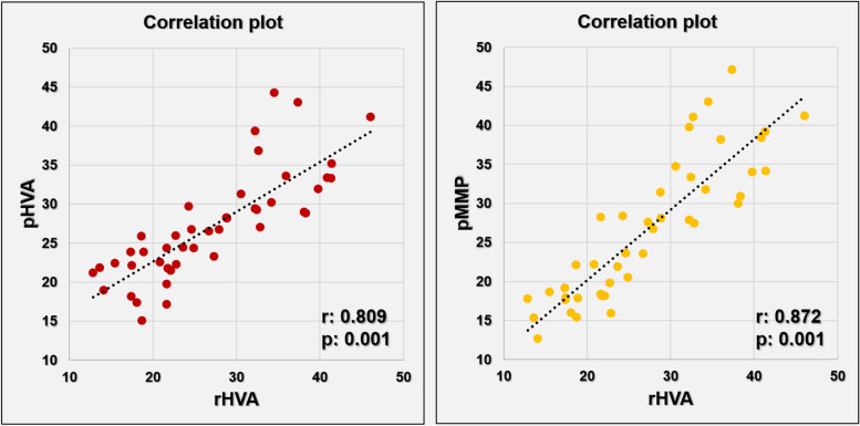
Results: The repeated rHVA, pHVA and pMMP measurements showed excellent intra and inter-observer reliability, with ICC values above 0.900. The mean rHVA, pHVA, and pMMP were statistically similar (p:0.929, 27.03°±8.7°, 27.11°±8.8° and 26.5°±9.0° respectively). The mean difference between the rHVA and pHVA was - 0.07°±5.1° (range, --9.67 to 9.56°), and the mean difference between the rHVA and pMMP was 0.53°±4.4° (range, -9.76° to 8.22°). There was a strong positive correlation between both photographic methods and radiographic measurements (rho = 0.809, p = 0.001 and rho = 0.872, p = 0.001). In the Bland Altman plot, the upper and lower LOAs (95%CI) ranged from - 10.11° to 9.93° for rHVA and pHVA, and from - 8.26° to 9.33° for rHVA and pMMP. Linear regression analysis showed a proportional bias for pHVA but not for the pMMP (p:0.010 versus p:0.633, respectively). The range of the mean difference (prediction interval) between the pMMP and rHVA was 17.59° and 20° for pHVA and rHVA. Simple linear regression showed that the rHVA was predicted by the following equation: rHVA = 4.73 + 0.84 × pMMP (r2 = 0.761, p < 0.001).
Conclusions: Although measuring HVA through smartphone photographs is reliable, it is not a valid prediction method.
Level of evidence: Level II, diagnostic assessment.
期刊介绍:
Journal of Foot and Ankle Research, the official journal of the Australian Podiatry Association and The College of Podiatry (UK), is an open access journal that encompasses all aspects of policy, organisation, delivery and clinical practice related to the assessment, diagnosis, prevention and management of foot and ankle disorders.
Journal of Foot and Ankle Research covers a wide range of clinical subject areas, including diabetology, paediatrics, sports medicine, gerontology and geriatrics, foot surgery, physical therapy, dermatology, wound management, radiology, biomechanics and bioengineering, orthotics and prosthetics, as well the broad areas of epidemiology, policy, organisation and delivery of services related to foot and ankle care.
The journal encourages submissions from all health professionals who manage lower limb conditions, including podiatrists, nurses, physical therapists and physiotherapists, orthopaedists, manual therapists, medical specialists and general medical practitioners, as well as health service researchers concerned with foot and ankle care.
The Australian Podiatry Association and the College of Podiatry (UK) have reserve funds to cover the article-processing charge for manuscripts submitted by its members. Society members can email the appropriate contact at Australian Podiatry Association or The College of Podiatry to obtain the corresponding code to enter on submission.


 分享
分享
 求助内容:
求助内容: 应助结果提醒方式:
 扫码关注我们
