Junxia Fu , Lvchen Cao , Shihui Wei , Ming Xu , Yali Song , Huiqi Li , Yuxia You
{"title":"一种基于gan的深度增强器,用于手持式眼底相机拍摄的视网膜图像的质量增强","authors":"Junxia Fu , Lvchen Cao , Shihui Wei , Ming Xu , Yali Song , Huiqi Li , Yuxia You","doi":"10.1016/j.aopr.2022.100077","DOIUrl":null,"url":null,"abstract":"<div><h3>Objective</h3><p>Due to limited imaging conditions, the quality of fundus images is often unsatisfactory, especially for images photographed by handheld fundus cameras. Here, we have developed an automated method based on combining two mirror-symmetric generative adversarial networks (GANs) for image enhancement.</p></div><div><h3>Methods</h3><p>A total of 1047 retinal images were included. The raw images were enhanced by a GAN-based deep enhancer and another methods based on luminosity and contrast adjustment. All raw images and enhanced images were anonymously assessed and classified into 6 levels of quality classification by three experienced ophthalmologists. The quality classification and quality change of images were compared. In addition, image-detailed reading results for the number of dubiously pathological fundi were also compared.</p></div><div><h3>Results</h3><p>After GAN enhancement, 42.9% of images increased their quality, 37.5% remained stable, and 19.6% decreased. After excluding the images at the highest level (level 0) before enhancement, a large number (75.6%) of images showed an increase in quality classification, and only a minority (9.3%) showed a decrease. The GAN-enhanced method was superior for quality improvement over a luminosity and contrast adjustment method (<em>P</em><0.001). In terms of image reading results, the consistency rate fluctuated from 86.6% to 95.6%, and for the specific disease subtypes, both discrepancy number and discrepancy rate were less than 15 and 15%, for two ophthalmologists.</p></div><div><h3>Conclusions</h3><p>Learning the style of high-quality retinal images based on the proposed deep enhancer may be an effective way to improve the quality of retinal images photographed by handheld fundus cameras.</p></div>","PeriodicalId":72103,"journal":{"name":"Advances in ophthalmology practice and research","volume":"2 3","pages":"Article 100077"},"PeriodicalIF":3.4000,"publicationDate":"2022-11-01","publicationTypes":"Journal Article","fieldsOfStudy":null,"isOpenAccess":false,"openAccessPdf":"https://ftp.ncbi.nlm.nih.gov/pub/pmc/oa_pdf/92/23/main.PMC10577846.pdf","citationCount":"2","resultStr":"{\"title\":\"A GAN-based deep enhancer for quality enhancement of retinal images photographed by a handheld fundus camera\",\"authors\":\"Junxia Fu , Lvchen Cao , Shihui Wei , Ming Xu , Yali Song , Huiqi Li , Yuxia You\",\"doi\":\"10.1016/j.aopr.2022.100077\",\"DOIUrl\":null,\"url\":null,\"abstract\":\"<div><h3>Objective</h3><p>Due to limited imaging conditions, the quality of fundus images is often unsatisfactory, especially for images photographed by handheld fundus cameras. Here, we have developed an automated method based on combining two mirror-symmetric generative adversarial networks (GANs) for image enhancement.</p></div><div><h3>Methods</h3><p>A total of 1047 retinal images were included. The raw images were enhanced by a GAN-based deep enhancer and another methods based on luminosity and contrast adjustment. All raw images and enhanced images were anonymously assessed and classified into 6 levels of quality classification by three experienced ophthalmologists. The quality classification and quality change of images were compared. In addition, image-detailed reading results for the number of dubiously pathological fundi were also compared.</p></div><div><h3>Results</h3><p>After GAN enhancement, 42.9% of images increased their quality, 37.5% remained stable, and 19.6% decreased. After excluding the images at the highest level (level 0) before enhancement, a large number (75.6%) of images showed an increase in quality classification, and only a minority (9.3%) showed a decrease. The GAN-enhanced method was superior for quality improvement over a luminosity and contrast adjustment method (<em>P</em><0.001). In terms of image reading results, the consistency rate fluctuated from 86.6% to 95.6%, and for the specific disease subtypes, both discrepancy number and discrepancy rate were less than 15 and 15%, for two ophthalmologists.</p></div><div><h3>Conclusions</h3><p>Learning the style of high-quality retinal images based on the proposed deep enhancer may be an effective way to improve the quality of retinal images photographed by handheld fundus cameras.</p></div>\",\"PeriodicalId\":72103,\"journal\":{\"name\":\"Advances in ophthalmology practice and research\",\"volume\":\"2 3\",\"pages\":\"Article 100077\"},\"PeriodicalIF\":3.4000,\"publicationDate\":\"2022-11-01\",\"publicationTypes\":\"Journal Article\",\"fieldsOfStudy\":null,\"isOpenAccess\":false,\"openAccessPdf\":\"https://ftp.ncbi.nlm.nih.gov/pub/pmc/oa_pdf/92/23/main.PMC10577846.pdf\",\"citationCount\":\"2\",\"resultStr\":null,\"platform\":\"Semanticscholar\",\"paperid\":null,\"PeriodicalName\":\"Advances in ophthalmology practice and research\",\"FirstCategoryId\":\"1085\",\"ListUrlMain\":\"https://www.sciencedirect.com/science/article/pii/S2667376222000543\",\"RegionNum\":0,\"RegionCategory\":null,\"ArticlePicture\":[],\"TitleCN\":null,\"AbstractTextCN\":null,\"PMCID\":null,\"EPubDate\":\"2022/8/19 0:00:00\",\"PubModel\":\"Epub\",\"JCR\":\"\",\"JCRName\":\"\",\"Score\":null,\"Total\":0}","platform":"Semanticscholar","paperid":null,"PeriodicalName":"Advances in ophthalmology practice and research","FirstCategoryId":"1085","ListUrlMain":"https://www.sciencedirect.com/science/article/pii/S2667376222000543","RegionNum":0,"RegionCategory":null,"ArticlePicture":[],"TitleCN":null,"AbstractTextCN":null,"PMCID":null,"EPubDate":"2022/8/19 0:00:00","PubModel":"Epub","JCR":"","JCRName":"","Score":null,"Total":0}
A GAN-based deep enhancer for quality enhancement of retinal images photographed by a handheld fundus camera
Objective
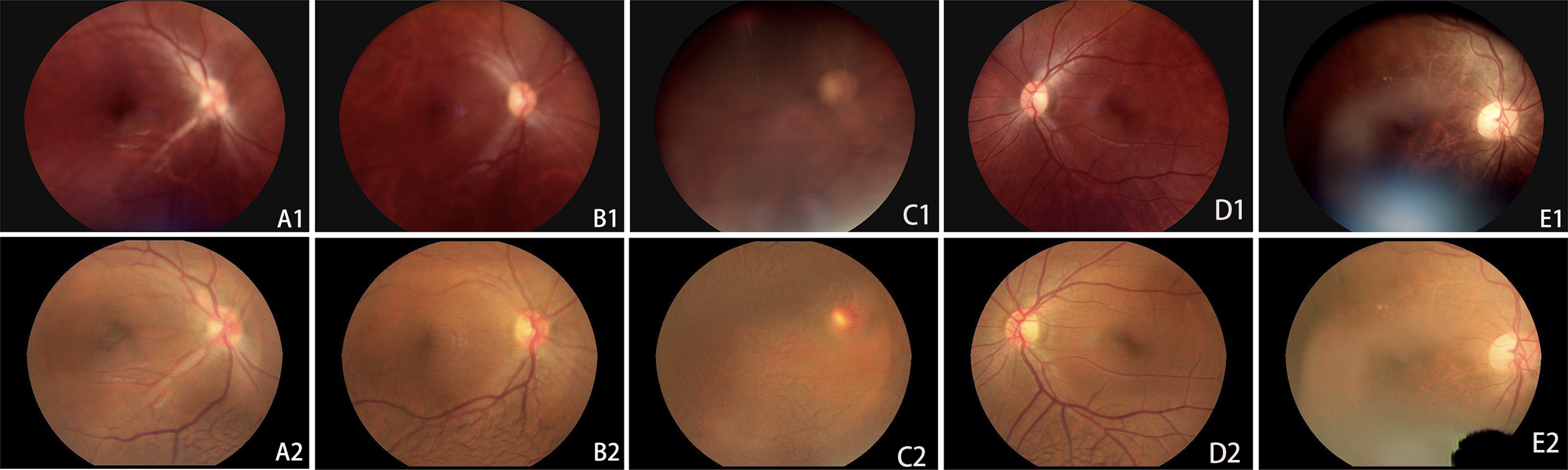
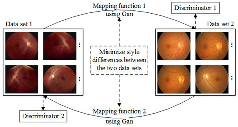
Due to limited imaging conditions, the quality of fundus images is often unsatisfactory, especially for images photographed by handheld fundus cameras. Here, we have developed an automated method based on combining two mirror-symmetric generative adversarial networks (GANs) for image enhancement.
Methods
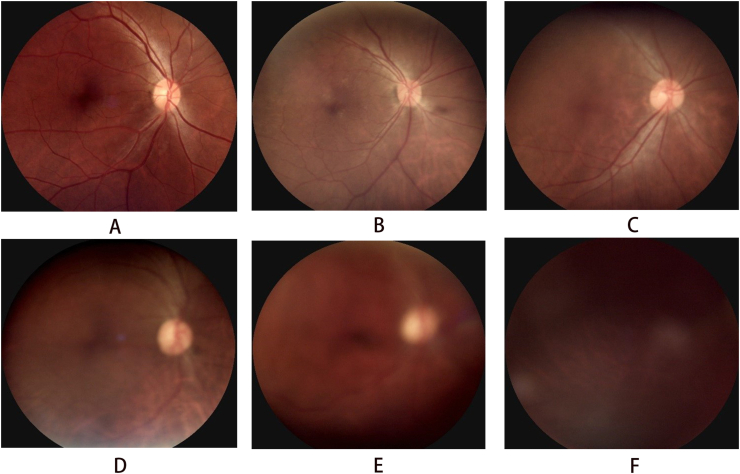
A total of 1047 retinal images were included. The raw images were enhanced by a GAN-based deep enhancer and another methods based on luminosity and contrast adjustment. All raw images and enhanced images were anonymously assessed and classified into 6 levels of quality classification by three experienced ophthalmologists. The quality classification and quality change of images were compared. In addition, image-detailed reading results for the number of dubiously pathological fundi were also compared.
Results
After GAN enhancement, 42.9% of images increased their quality, 37.5% remained stable, and 19.6% decreased. After excluding the images at the highest level (level 0) before enhancement, a large number (75.6%) of images showed an increase in quality classification, and only a minority (9.3%) showed a decrease. The GAN-enhanced method was superior for quality improvement over a luminosity and contrast adjustment method (P<0.001). In terms of image reading results, the consistency rate fluctuated from 86.6% to 95.6%, and for the specific disease subtypes, both discrepancy number and discrepancy rate were less than 15 and 15%, for two ophthalmologists.
Conclusions
Learning the style of high-quality retinal images based on the proposed deep enhancer may be an effective way to improve the quality of retinal images photographed by handheld fundus cameras.


 分享
分享
 求助内容:
求助内容: 应助结果提醒方式:
 扫码关注我们
