Brittany M Stopa, James Crowley, Csaba Juhász, Cara M Rogers, Mark R Witcher, Jackson W Kiser
{"title":"前列腺特异性膜抗原作为中枢神经系统肿瘤神经成像的靶点","authors":"Brittany M Stopa, James Crowley, Csaba Juhász, Cara M Rogers, Mark R Witcher, Jackson W Kiser","doi":"10.1155/2022/5358545","DOIUrl":null,"url":null,"abstract":"<p><strong>Introduction: </strong>Positron emission tomography (PET) imaging with prostate-specific membrane antigen- (PSMA-) binding tracers has been found incidentally to demonstrate uptake in CNS tumors. Following the encouraging findings of several such case reports, there is a growing interest in the potential application of PSMA-targeted PET imaging for diagnostics, theranostics, and monitoring of CNS tumors. This is a systematic literature review on PSMA-binding tracers in CNS tumors.</p><p><strong>Methods: </strong>A PubMed search was conducted, including preclinical and clinical reports. One hundred and twelve records were identified, and after screening, 56 were included in the final report.</p><p><strong>Results: </strong>Tissue studies demonstrated PSMA expression in tumor vascular endothelial cells, without expression in normal brain tissue, though the extent and intensity of staining varied by anti-PSMA antibody and methodology. Most included studies reported on gliomas, which showed strong PSMA ligand uptake and more favorable tumor to background ratios than other PET tracers. There are also case reports demonstrating PSMA ligand uptake in prostate cancer brain metastases, nonprostate cancer brain metastases, and meningiomas. We also review the properties of the various PSMA-binding radiotracers available. Therapeutic and theranostic applications of PSMA-binding tracers have been studied, including labeled alpha- and beta-ray emitting isotopes, as well as PSMA targeting in directing MRI-guided focused ultrasound.</p><p><strong>Conclusions: </strong>There is a potential application for PSMA-targeted PET in neuro-oncology as a combination of diagnostic and therapeutic use, as a theranostic modality for managing CNS tumors. Further research is needed regarding the mechanism(s) of PSMA expression in CNS tumors and its differential performance by tumor type.</p>","PeriodicalId":49796,"journal":{"name":"Molecular Imaging","volume":"2022 1","pages":"5358545"},"PeriodicalIF":2.4000,"publicationDate":"2022-04-15","publicationTypes":"Journal Article","fieldsOfStudy":null,"isOpenAccess":false,"openAccessPdf":"https://www.ncbi.nlm.nih.gov/pmc/articles/PMC9042374/pdf/","citationCount":"0","resultStr":"{\"title\":\"Prostate-Specific Membrane Antigen as Target for Neuroimaging of Central Nervous System Tumors.\",\"authors\":\"Brittany M Stopa, James Crowley, Csaba Juhász, Cara M Rogers, Mark R Witcher, Jackson W Kiser\",\"doi\":\"10.1155/2022/5358545\",\"DOIUrl\":null,\"url\":null,\"abstract\":\"<p><strong>Introduction: </strong>Positron emission tomography (PET) imaging with prostate-specific membrane antigen- (PSMA-) binding tracers has been found incidentally to demonstrate uptake in CNS tumors. Following the encouraging findings of several such case reports, there is a growing interest in the potential application of PSMA-targeted PET imaging for diagnostics, theranostics, and monitoring of CNS tumors. This is a systematic literature review on PSMA-binding tracers in CNS tumors.</p><p><strong>Methods: </strong>A PubMed search was conducted, including preclinical and clinical reports. One hundred and twelve records were identified, and after screening, 56 were included in the final report.</p><p><strong>Results: </strong>Tissue studies demonstrated PSMA expression in tumor vascular endothelial cells, without expression in normal brain tissue, though the extent and intensity of staining varied by anti-PSMA antibody and methodology. Most included studies reported on gliomas, which showed strong PSMA ligand uptake and more favorable tumor to background ratios than other PET tracers. There are also case reports demonstrating PSMA ligand uptake in prostate cancer brain metastases, nonprostate cancer brain metastases, and meningiomas. We also review the properties of the various PSMA-binding radiotracers available. Therapeutic and theranostic applications of PSMA-binding tracers have been studied, including labeled alpha- and beta-ray emitting isotopes, as well as PSMA targeting in directing MRI-guided focused ultrasound.</p><p><strong>Conclusions: </strong>There is a potential application for PSMA-targeted PET in neuro-oncology as a combination of diagnostic and therapeutic use, as a theranostic modality for managing CNS tumors. Further research is needed regarding the mechanism(s) of PSMA expression in CNS tumors and its differential performance by tumor type.</p>\",\"PeriodicalId\":49796,\"journal\":{\"name\":\"Molecular Imaging\",\"volume\":\"2022 1\",\"pages\":\"5358545\"},\"PeriodicalIF\":2.4000,\"publicationDate\":\"2022-04-15\",\"publicationTypes\":\"Journal Article\",\"fieldsOfStudy\":null,\"isOpenAccess\":false,\"openAccessPdf\":\"https://www.ncbi.nlm.nih.gov/pmc/articles/PMC9042374/pdf/\",\"citationCount\":\"0\",\"resultStr\":null,\"platform\":\"Semanticscholar\",\"paperid\":null,\"PeriodicalName\":\"Molecular Imaging\",\"FirstCategoryId\":\"3\",\"ListUrlMain\":\"https://doi.org/10.1155/2022/5358545\",\"RegionNum\":4,\"RegionCategory\":\"医学\",\"ArticlePicture\":[],\"TitleCN\":null,\"AbstractTextCN\":null,\"PMCID\":null,\"EPubDate\":\"2022/1/1 0:00:00\",\"PubModel\":\"eCollection\",\"JCR\":\"Q2\",\"JCRName\":\"Medicine\",\"Score\":null,\"Total\":0}","platform":"Semanticscholar","paperid":null,"PeriodicalName":"Molecular Imaging","FirstCategoryId":"3","ListUrlMain":"https://doi.org/10.1155/2022/5358545","RegionNum":4,"RegionCategory":"医学","ArticlePicture":[],"TitleCN":null,"AbstractTextCN":null,"PMCID":null,"EPubDate":"2022/1/1 0:00:00","PubModel":"eCollection","JCR":"Q2","JCRName":"Medicine","Score":null,"Total":0}
Prostate-Specific Membrane Antigen as Target for Neuroimaging of Central Nervous System Tumors.
Introduction: Positron emission tomography (PET) imaging with prostate-specific membrane antigen- (PSMA-) binding tracers has been found incidentally to demonstrate uptake in CNS tumors. Following the encouraging findings of several such case reports, there is a growing interest in the potential application of PSMA-targeted PET imaging for diagnostics, theranostics, and monitoring of CNS tumors. This is a systematic literature review on PSMA-binding tracers in CNS tumors.
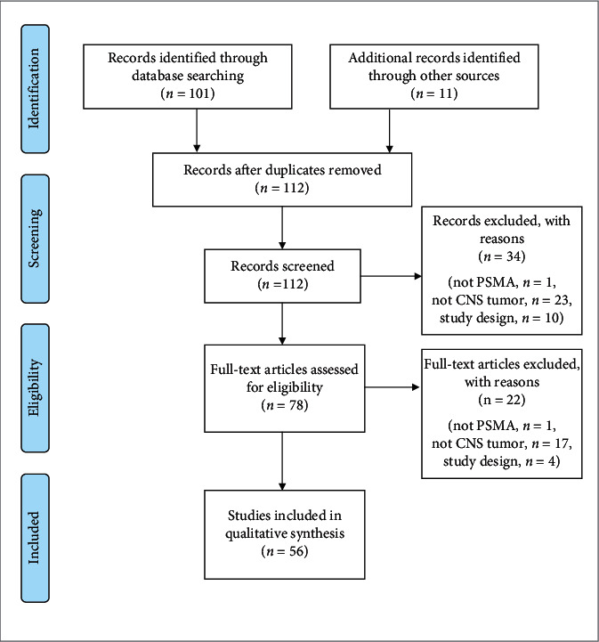
Methods: A PubMed search was conducted, including preclinical and clinical reports. One hundred and twelve records were identified, and after screening, 56 were included in the final report.
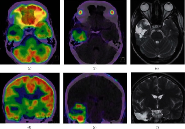
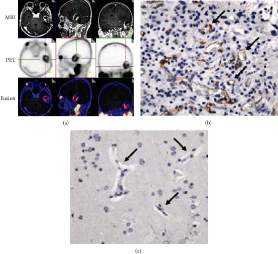
Results: Tissue studies demonstrated PSMA expression in tumor vascular endothelial cells, without expression in normal brain tissue, though the extent and intensity of staining varied by anti-PSMA antibody and methodology. Most included studies reported on gliomas, which showed strong PSMA ligand uptake and more favorable tumor to background ratios than other PET tracers. There are also case reports demonstrating PSMA ligand uptake in prostate cancer brain metastases, nonprostate cancer brain metastases, and meningiomas. We also review the properties of the various PSMA-binding radiotracers available. Therapeutic and theranostic applications of PSMA-binding tracers have been studied, including labeled alpha- and beta-ray emitting isotopes, as well as PSMA targeting in directing MRI-guided focused ultrasound.
Conclusions: There is a potential application for PSMA-targeted PET in neuro-oncology as a combination of diagnostic and therapeutic use, as a theranostic modality for managing CNS tumors. Further research is needed regarding the mechanism(s) of PSMA expression in CNS tumors and its differential performance by tumor type.
期刊介绍:
Molecular Imaging is a peer-reviewed, open access journal highlighting the breadth of molecular imaging research from basic science to preclinical studies to human applications. This serves both the scientific and clinical communities by disseminating novel results and concepts relevant to the biological study of normal and disease processes in both basic and translational studies ranging from mice to humans.


 分享
分享
 求助内容:
求助内容: 应助结果提醒方式:
 扫码关注我们
