Fernando Bandeira Sulczewski , Larissa Alves Martino , Bianca da Silva Almeida , Márcio Massao Yamamoto , Daniela Santoro Rosa , Silvia Beatriz Boscardin
{"title":"STAT6信号通路控制抗原靶向常规2型树突状细胞后促进的生发中心反应","authors":"Fernando Bandeira Sulczewski , Larissa Alves Martino , Bianca da Silva Almeida , Márcio Massao Yamamoto , Daniela Santoro Rosa , Silvia Beatriz Boscardin","doi":"10.1016/j.crimmu.2021.08.001","DOIUrl":null,"url":null,"abstract":"<div><p>Conventional dendritic cells (cDCs) are antigen-presenting cells specialized in naïve T cell priming. Mice splenic cDCs are classified as cDC1s and cDC2s, and their main functions have been elucidated in the last decade. While cDC1s are specialized in priming type 1 helper T cells (T<sub>H</sub>1) and in cross presentation, cDC2s prime T follicular helper (T<sub>FH</sub>) cells that stimulate germinal center (GC) formation, plasma cell differentiation and antibody production. However, less is known about the molecular mechanisms used by cDCs to prime those responses. Here, using WT and STAT6-deficient mice (STAT6 KO), we targeted a model antigen to cDC1s and cDC2s via DEC205 and DCIR2 receptors, respectively, in an attempt to study whether the STAT6 signaling pathway would modulate cDCs’ ability to prime helper T cells. We show that the differentiation and maturation of cDCs, after stimulation with an adjuvant, were comparable between WT and STAT6 KO mice. Besides, our results indicate that, in STAT6 KO mice, antigen targeting to cDC2s induced reduced T<sub>FH</sub> and GC responses, but did not alter plasma cells numbers and antibody titers. Thus, we conclude that the STAT6 signaling pathway modulates the immune response after antigen targeting to cDC2s via the DCIR2 receptor: while STAT6 stimulates the development of T<sub>FH</sub> cells and GC formation, plasma cell differentiation occurs in a STAT6 independent manner.</p></div>","PeriodicalId":72750,"journal":{"name":"Current research in immunology","volume":"2 ","pages":"Pages 120-131"},"PeriodicalIF":0.0000,"publicationDate":"2021-01-01","publicationTypes":"Journal Article","fieldsOfStudy":null,"isOpenAccess":false,"openAccessPdf":"https://sci-hub-pdf.com/10.1016/j.crimmu.2021.08.001","citationCount":"4","resultStr":"{\"title\":\"STAT6 signaling pathway controls germinal center responses promoted after antigen targeting to conventional type 2 dendritic cells\",\"authors\":\"Fernando Bandeira Sulczewski , Larissa Alves Martino , Bianca da Silva Almeida , Márcio Massao Yamamoto , Daniela Santoro Rosa , Silvia Beatriz Boscardin\",\"doi\":\"10.1016/j.crimmu.2021.08.001\",\"DOIUrl\":null,\"url\":null,\"abstract\":\"<div><p>Conventional dendritic cells (cDCs) are antigen-presenting cells specialized in naïve T cell priming. Mice splenic cDCs are classified as cDC1s and cDC2s, and their main functions have been elucidated in the last decade. While cDC1s are specialized in priming type 1 helper T cells (T<sub>H</sub>1) and in cross presentation, cDC2s prime T follicular helper (T<sub>FH</sub>) cells that stimulate germinal center (GC) formation, plasma cell differentiation and antibody production. However, less is known about the molecular mechanisms used by cDCs to prime those responses. Here, using WT and STAT6-deficient mice (STAT6 KO), we targeted a model antigen to cDC1s and cDC2s via DEC205 and DCIR2 receptors, respectively, in an attempt to study whether the STAT6 signaling pathway would modulate cDCs’ ability to prime helper T cells. We show that the differentiation and maturation of cDCs, after stimulation with an adjuvant, were comparable between WT and STAT6 KO mice. Besides, our results indicate that, in STAT6 KO mice, antigen targeting to cDC2s induced reduced T<sub>FH</sub> and GC responses, but did not alter plasma cells numbers and antibody titers. Thus, we conclude that the STAT6 signaling pathway modulates the immune response after antigen targeting to cDC2s via the DCIR2 receptor: while STAT6 stimulates the development of T<sub>FH</sub> cells and GC formation, plasma cell differentiation occurs in a STAT6 independent manner.</p></div>\",\"PeriodicalId\":72750,\"journal\":{\"name\":\"Current research in immunology\",\"volume\":\"2 \",\"pages\":\"Pages 120-131\"},\"PeriodicalIF\":0.0000,\"publicationDate\":\"2021-01-01\",\"publicationTypes\":\"Journal Article\",\"fieldsOfStudy\":null,\"isOpenAccess\":false,\"openAccessPdf\":\"https://sci-hub-pdf.com/10.1016/j.crimmu.2021.08.001\",\"citationCount\":\"4\",\"resultStr\":null,\"platform\":\"Semanticscholar\",\"paperid\":null,\"PeriodicalName\":\"Current research in immunology\",\"FirstCategoryId\":\"1085\",\"ListUrlMain\":\"https://www.sciencedirect.com/science/article/pii/S2590255521000135\",\"RegionNum\":0,\"RegionCategory\":null,\"ArticlePicture\":[],\"TitleCN\":null,\"AbstractTextCN\":null,\"PMCID\":null,\"EPubDate\":\"2021/8/28 0:00:00\",\"PubModel\":\"Epub\",\"JCR\":\"Q4\",\"JCRName\":\"Immunology and Microbiology\",\"Score\":null,\"Total\":0}","platform":"Semanticscholar","paperid":null,"PeriodicalName":"Current research in immunology","FirstCategoryId":"1085","ListUrlMain":"https://www.sciencedirect.com/science/article/pii/S2590255521000135","RegionNum":0,"RegionCategory":null,"ArticlePicture":[],"TitleCN":null,"AbstractTextCN":null,"PMCID":null,"EPubDate":"2021/8/28 0:00:00","PubModel":"Epub","JCR":"Q4","JCRName":"Immunology and Microbiology","Score":null,"Total":0}
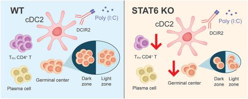
STAT6 signaling pathway controls germinal center responses promoted after antigen targeting to conventional type 2 dendritic cells
Conventional dendritic cells (cDCs) are antigen-presenting cells specialized in naïve T cell priming. Mice splenic cDCs are classified as cDC1s and cDC2s, and their main functions have been elucidated in the last decade. While cDC1s are specialized in priming type 1 helper T cells (TH1) and in cross presentation, cDC2s prime T follicular helper (TFH) cells that stimulate germinal center (GC) formation, plasma cell differentiation and antibody production. However, less is known about the molecular mechanisms used by cDCs to prime those responses. Here, using WT and STAT6-deficient mice (STAT6 KO), we targeted a model antigen to cDC1s and cDC2s via DEC205 and DCIR2 receptors, respectively, in an attempt to study whether the STAT6 signaling pathway would modulate cDCs’ ability to prime helper T cells. We show that the differentiation and maturation of cDCs, after stimulation with an adjuvant, were comparable between WT and STAT6 KO mice. Besides, our results indicate that, in STAT6 KO mice, antigen targeting to cDC2s induced reduced TFH and GC responses, but did not alter plasma cells numbers and antibody titers. Thus, we conclude that the STAT6 signaling pathway modulates the immune response after antigen targeting to cDC2s via the DCIR2 receptor: while STAT6 stimulates the development of TFH cells and GC formation, plasma cell differentiation occurs in a STAT6 independent manner.


 分享
分享
 求助内容:
求助内容: 应助结果提醒方式:
 扫码关注我们
