{"title":"1例严重急性呼吸综合征冠状病毒患者鼻内镜图像的光学分析","authors":"Sujeong Bae, Young Joon Jun","doi":"10.18787/jr.2022.00402","DOIUrl":null,"url":null,"abstract":"<p><p>Severe acute respiratory syndrome coronavirus 2 (SARS-CoV-2) has spread worldwide; a simple diagnostic test is becoming increasingly important. Polymerase chain reaction tests of nasal swab samples are expensive and time-consuming. Detailed observation of the nasal cavity is required. No nasal endoscopic study regarding patients with SARS-CoV-2 has been published. Here, we compared endoscopic images of normal subjects and patients with allergic rhinitis (AR) to images from a patient with confirmed SARS-CoV-2 infection. Nasal cavity endoscopic images (of the inferior turbinate) were compared using the hue, saturation, and luminance (HSL) color system of Matrox Design Assistant, version 5.0. The multiple allergen simultaneous test was used to distinguish normal subjects from patients with AR. HSL patterns were compared among images. We enrolled 14 normal subjects and patients with AR and 1 patient with confirmed SARS-CoV-2 infection. The inferior turbinates of normal subjects and patients with AR were smooth and uniform, while those of the patient with SARS-CoV-2 were scratched and stretched. Although additional data are needed, optical analysis of nasal endoscopy images may be useful to screen for SARS-CoV-2 infection. This imaging method is rapid, non-invasive, and inexpensive.</p>","PeriodicalId":33935,"journal":{"name":"Journal of Rhinology","volume":" ","pages":"96-100"},"PeriodicalIF":0.0000,"publicationDate":"2022-07-01","publicationTypes":"Journal Article","fieldsOfStudy":null,"isOpenAccess":false,"openAccessPdf":"https://www.ncbi.nlm.nih.gov/pmc/articles/PMC11540236/pdf/","citationCount":"0","resultStr":"{\"title\":\"Optical Analysis of Nasal Endoscopic Images From a Patient With Severe Acute Respiratory Syndrome Coronavirus 2.\",\"authors\":\"Sujeong Bae, Young Joon Jun\",\"doi\":\"10.18787/jr.2022.00402\",\"DOIUrl\":null,\"url\":null,\"abstract\":\"<p><p>Severe acute respiratory syndrome coronavirus 2 (SARS-CoV-2) has spread worldwide; a simple diagnostic test is becoming increasingly important. Polymerase chain reaction tests of nasal swab samples are expensive and time-consuming. Detailed observation of the nasal cavity is required. No nasal endoscopic study regarding patients with SARS-CoV-2 has been published. Here, we compared endoscopic images of normal subjects and patients with allergic rhinitis (AR) to images from a patient with confirmed SARS-CoV-2 infection. Nasal cavity endoscopic images (of the inferior turbinate) were compared using the hue, saturation, and luminance (HSL) color system of Matrox Design Assistant, version 5.0. The multiple allergen simultaneous test was used to distinguish normal subjects from patients with AR. HSL patterns were compared among images. We enrolled 14 normal subjects and patients with AR and 1 patient with confirmed SARS-CoV-2 infection. The inferior turbinates of normal subjects and patients with AR were smooth and uniform, while those of the patient with SARS-CoV-2 were scratched and stretched. Although additional data are needed, optical analysis of nasal endoscopy images may be useful to screen for SARS-CoV-2 infection. This imaging method is rapid, non-invasive, and inexpensive.</p>\",\"PeriodicalId\":33935,\"journal\":{\"name\":\"Journal of Rhinology\",\"volume\":\" \",\"pages\":\"96-100\"},\"PeriodicalIF\":0.0000,\"publicationDate\":\"2022-07-01\",\"publicationTypes\":\"Journal Article\",\"fieldsOfStudy\":null,\"isOpenAccess\":false,\"openAccessPdf\":\"https://www.ncbi.nlm.nih.gov/pmc/articles/PMC11540236/pdf/\",\"citationCount\":\"0\",\"resultStr\":null,\"platform\":\"Semanticscholar\",\"paperid\":null,\"PeriodicalName\":\"Journal of Rhinology\",\"FirstCategoryId\":\"1085\",\"ListUrlMain\":\"https://doi.org/10.18787/jr.2022.00402\",\"RegionNum\":0,\"RegionCategory\":null,\"ArticlePicture\":[],\"TitleCN\":null,\"AbstractTextCN\":null,\"PMCID\":null,\"EPubDate\":\"2022/7/27 0:00:00\",\"PubModel\":\"Epub\",\"JCR\":\"\",\"JCRName\":\"\",\"Score\":null,\"Total\":0}","platform":"Semanticscholar","paperid":null,"PeriodicalName":"Journal of Rhinology","FirstCategoryId":"1085","ListUrlMain":"https://doi.org/10.18787/jr.2022.00402","RegionNum":0,"RegionCategory":null,"ArticlePicture":[],"TitleCN":null,"AbstractTextCN":null,"PMCID":null,"EPubDate":"2022/7/27 0:00:00","PubModel":"Epub","JCR":"","JCRName":"","Score":null,"Total":0}
Optical Analysis of Nasal Endoscopic Images From a Patient With Severe Acute Respiratory Syndrome Coronavirus 2.
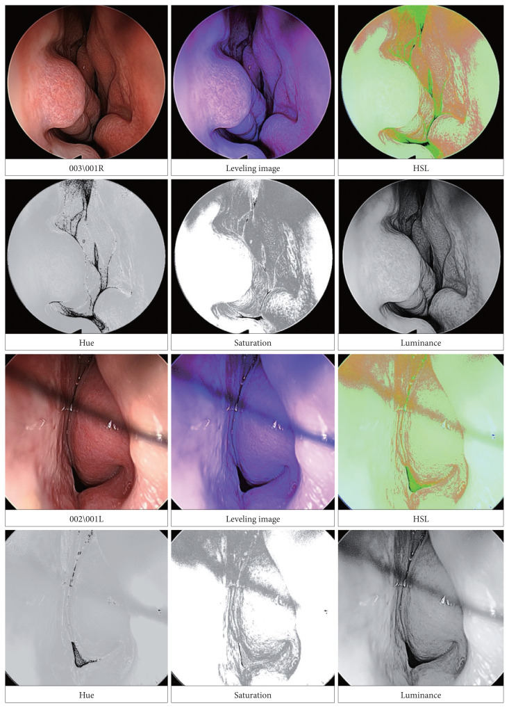
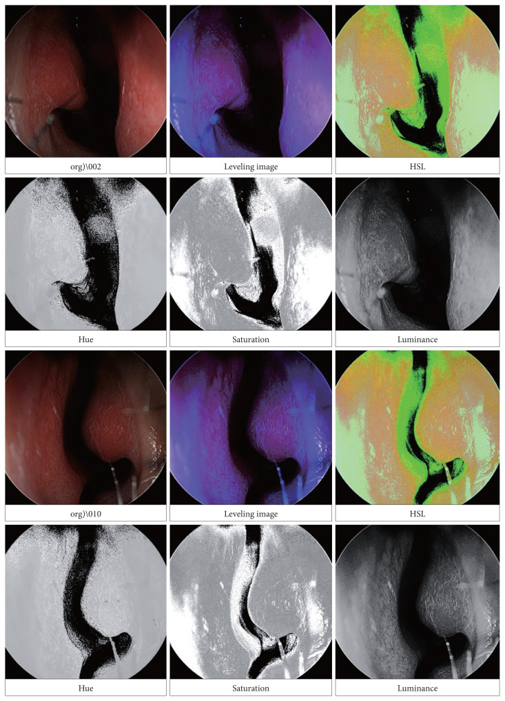
Severe acute respiratory syndrome coronavirus 2 (SARS-CoV-2) has spread worldwide; a simple diagnostic test is becoming increasingly important. Polymerase chain reaction tests of nasal swab samples are expensive and time-consuming. Detailed observation of the nasal cavity is required. No nasal endoscopic study regarding patients with SARS-CoV-2 has been published. Here, we compared endoscopic images of normal subjects and patients with allergic rhinitis (AR) to images from a patient with confirmed SARS-CoV-2 infection. Nasal cavity endoscopic images (of the inferior turbinate) were compared using the hue, saturation, and luminance (HSL) color system of Matrox Design Assistant, version 5.0. The multiple allergen simultaneous test was used to distinguish normal subjects from patients with AR. HSL patterns were compared among images. We enrolled 14 normal subjects and patients with AR and 1 patient with confirmed SARS-CoV-2 infection. The inferior turbinates of normal subjects and patients with AR were smooth and uniform, while those of the patient with SARS-CoV-2 were scratched and stretched. Although additional data are needed, optical analysis of nasal endoscopy images may be useful to screen for SARS-CoV-2 infection. This imaging method is rapid, non-invasive, and inexpensive.


 分享
分享

 求助内容:
求助内容: 应助结果提醒方式:
 扫码关注我们
