IF 1.4 3区 医学Q4 BIOCHEMISTRY & MOLECULAR BIOLOGYMolecular VisionPub Date : 2023-07-16eCollection Date: 2023-01-01
Chase Paterson, Jamen Cannon, Elizabeth Vargis
{"title":"早期RPE细胞连接缺失对体外VEGF、Ang-2和TIMP分泌的影响。","authors":"Chase Paterson, Jamen Cannon, Elizabeth Vargis","doi":"","DOIUrl":null,"url":null,"abstract":"<p><strong>Purpose: </strong>The retinal pigment epithelium (RPE) is an important tissue for maintaining a healthy retina. Retinal pigment epithelial cells help regulate nutrient transport to photoreceptors and are heavily pigmented to prevent light scattering. These cells also have junction proteins to form monolayers. Monolayers are key players in pathologies such as age-related macular degeneration (AMD), a leading cause of vision loss in older adults. During AMD, RPE cell detachment can occur, resulting in a loss of junctions. Losing junctions can increase the expression of pro-angiogenic vascular endothelial growth factor (VEGF). This overexpression can cause abnormal blood vessel growth or angiogenesis in the retina. Age-related macular degeneration treatments target VEGF to slow angiogenesis progression. However, other proteins, such as angiopoietin-2 (Ang-2) and the tissue inhibitor of metalloproteinase-1 (TIMP-1), may also play important roles, making them potential targets for treatment. Controlling RPE junction formation will help elucidate the relationship between RPE cell detachment and additional angiogenic factor secretion, lead to more therapeutics, and increase the efficacy of current treatments.</p><p><strong>Methods: </strong>Micropatterning was used to control the spatial arrangement of primary porcine RPE cells using polydimethylsiloxane (PDMS) stencils. Patterns were formed into PDMS stencils to mimic 10%, 25%, and 50% overall detachment of the RPE monolayer. Zonula-occludens-1 (ZO-1), Ang-2, and VEGF were visualized using immunocytochemical (ICC) staining. An enzyme-linked immunosorbent assay (ELISA) was used to quantify extracellular Ang-2, VEGF, TIMP-1, and TIMP-2 levels. A rod outer segment (OS) phagocytosis assay was performed to determine how RPE junction loss directly affects photoreceptor support.</p><p><strong>Results: </strong>The growth of primary porcine RPE cells was successfully controlled using stencils. Morphological changes and a decrease in pigmentation were observed, showing a decline in barrier and light absorption functions as degeneration increased. One day after stencil removal, junction proteins were delocalized, and angiogenic factor secretions were correlated with increased levels of detachment. Secretion levels of Ang-2 and TIMP-1 were significantly increased, whereas VEGF and TIMP-2 concentrations were not as affected by varying levels of detachment. OS phagocytosis appeared lower in RPE cells when ZO-1 was affected.</p><p><strong>Conclusions: </strong>These results suggest a correlation between loss of junctions, abnormal angiogenic protein secretion, and reduced OS phagocytosis. Furthermore, Ang-2 and TIMP-1 proteins might be beneficial targets for AMD treatments, and their roles in retinal diseases deserve further investigation.</p>","PeriodicalId":18866,"journal":{"name":"Molecular Vision","volume":"29 ","pages":"87-101"},"PeriodicalIF":1.4000,"publicationDate":"2023-07-16","publicationTypes":"Journal Article","fieldsOfStudy":null,"isOpenAccess":false,"openAccessPdf":"https://ftp.ncbi.nlm.nih.gov/pub/pmc/oa_pdf/d8/e0/mv-v29-87.PMC10584031.pdf","citationCount":"0","resultStr":"{\"title\":\"The impact of early RPE cell junction loss on VEGF, Ang-2, and TIMP secretion in vitro.\",\"authors\":\"Chase Paterson, Jamen Cannon, Elizabeth Vargis\",\"doi\":\"\",\"DOIUrl\":null,\"url\":null,\"abstract\":\"<p><strong>Purpose: </strong>The retinal pigment epithelium (RPE) is an important tissue for maintaining a healthy retina. Retinal pigment epithelial cells help regulate nutrient transport to photoreceptors and are heavily pigmented to prevent light scattering. These cells also have junction proteins to form monolayers. Monolayers are key players in pathologies such as age-related macular degeneration (AMD), a leading cause of vision loss in older adults. During AMD, RPE cell detachment can occur, resulting in a loss of junctions. Losing junctions can increase the expression of pro-angiogenic vascular endothelial growth factor (VEGF). This overexpression can cause abnormal blood vessel growth or angiogenesis in the retina. Age-related macular degeneration treatments target VEGF to slow angiogenesis progression. However, other proteins, such as angiopoietin-2 (Ang-2) and the tissue inhibitor of metalloproteinase-1 (TIMP-1), may also play important roles, making them potential targets for treatment. Controlling RPE junction formation will help elucidate the relationship between RPE cell detachment and additional angiogenic factor secretion, lead to more therapeutics, and increase the efficacy of current treatments.</p><p><strong>Methods: </strong>Micropatterning was used to control the spatial arrangement of primary porcine RPE cells using polydimethylsiloxane (PDMS) stencils. Patterns were formed into PDMS stencils to mimic 10%, 25%, and 50% overall detachment of the RPE monolayer. Zonula-occludens-1 (ZO-1), Ang-2, and VEGF were visualized using immunocytochemical (ICC) staining. An enzyme-linked immunosorbent assay (ELISA) was used to quantify extracellular Ang-2, VEGF, TIMP-1, and TIMP-2 levels. A rod outer segment (OS) phagocytosis assay was performed to determine how RPE junction loss directly affects photoreceptor support.</p><p><strong>Results: </strong>The growth of primary porcine RPE cells was successfully controlled using stencils. Morphological changes and a decrease in pigmentation were observed, showing a decline in barrier and light absorption functions as degeneration increased. One day after stencil removal, junction proteins were delocalized, and angiogenic factor secretions were correlated with increased levels of detachment. Secretion levels of Ang-2 and TIMP-1 were significantly increased, whereas VEGF and TIMP-2 concentrations were not as affected by varying levels of detachment. OS phagocytosis appeared lower in RPE cells when ZO-1 was affected.</p><p><strong>Conclusions: </strong>These results suggest a correlation between loss of junctions, abnormal angiogenic protein secretion, and reduced OS phagocytosis. Furthermore, Ang-2 and TIMP-1 proteins might be beneficial targets for AMD treatments, and their roles in retinal diseases deserve further investigation.</p>\",\"PeriodicalId\":18866,\"journal\":{\"name\":\"Molecular Vision\",\"volume\":\"29 \",\"pages\":\"87-101\"},\"PeriodicalIF\":1.4000,\"publicationDate\":\"2023-07-16\",\"publicationTypes\":\"Journal Article\",\"fieldsOfStudy\":null,\"isOpenAccess\":false,\"openAccessPdf\":\"https://ftp.ncbi.nlm.nih.gov/pub/pmc/oa_pdf/d8/e0/mv-v29-87.PMC10584031.pdf\",\"citationCount\":\"0\",\"resultStr\":null,\"platform\":\"Semanticscholar\",\"paperid\":null,\"PeriodicalName\":\"Molecular Vision\",\"FirstCategoryId\":\"3\",\"ListUrlMain\":\"\",\"RegionNum\":3,\"RegionCategory\":\"医学\",\"ArticlePicture\":[],\"TitleCN\":null,\"AbstractTextCN\":null,\"PMCID\":null,\"EPubDate\":\"2023/1/1 0:00:00\",\"PubModel\":\"eCollection\",\"JCR\":\"Q4\",\"JCRName\":\"BIOCHEMISTRY & MOLECULAR BIOLOGY\",\"Score\":null,\"Total\":0}","platform":"Semanticscholar","paperid":null,"PeriodicalName":"Molecular Vision","FirstCategoryId":"3","ListUrlMain":"","RegionNum":3,"RegionCategory":"医学","ArticlePicture":[],"TitleCN":null,"AbstractTextCN":null,"PMCID":null,"EPubDate":"2023/1/1 0:00:00","PubModel":"eCollection","JCR":"Q4","JCRName":"BIOCHEMISTRY & MOLECULAR BIOLOGY","Score":null,"Total":0}
The impact of early RPE cell junction loss on VEGF, Ang-2, and TIMP secretion in vitro.
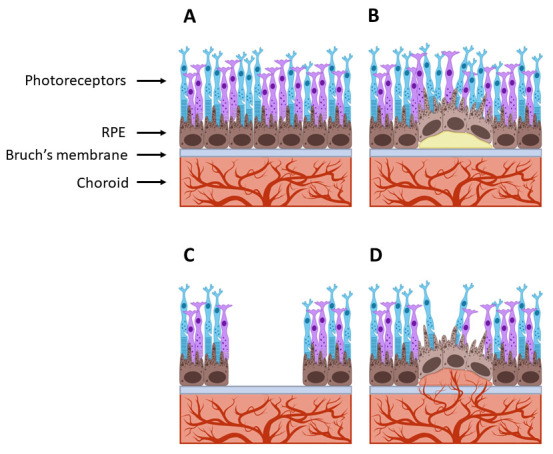
Purpose: The retinal pigment epithelium (RPE) is an important tissue for maintaining a healthy retina. Retinal pigment epithelial cells help regulate nutrient transport to photoreceptors and are heavily pigmented to prevent light scattering. These cells also have junction proteins to form monolayers. Monolayers are key players in pathologies such as age-related macular degeneration (AMD), a leading cause of vision loss in older adults. During AMD, RPE cell detachment can occur, resulting in a loss of junctions. Losing junctions can increase the expression of pro-angiogenic vascular endothelial growth factor (VEGF). This overexpression can cause abnormal blood vessel growth or angiogenesis in the retina. Age-related macular degeneration treatments target VEGF to slow angiogenesis progression. However, other proteins, such as angiopoietin-2 (Ang-2) and the tissue inhibitor of metalloproteinase-1 (TIMP-1), may also play important roles, making them potential targets for treatment. Controlling RPE junction formation will help elucidate the relationship between RPE cell detachment and additional angiogenic factor secretion, lead to more therapeutics, and increase the efficacy of current treatments.
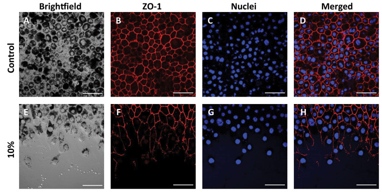
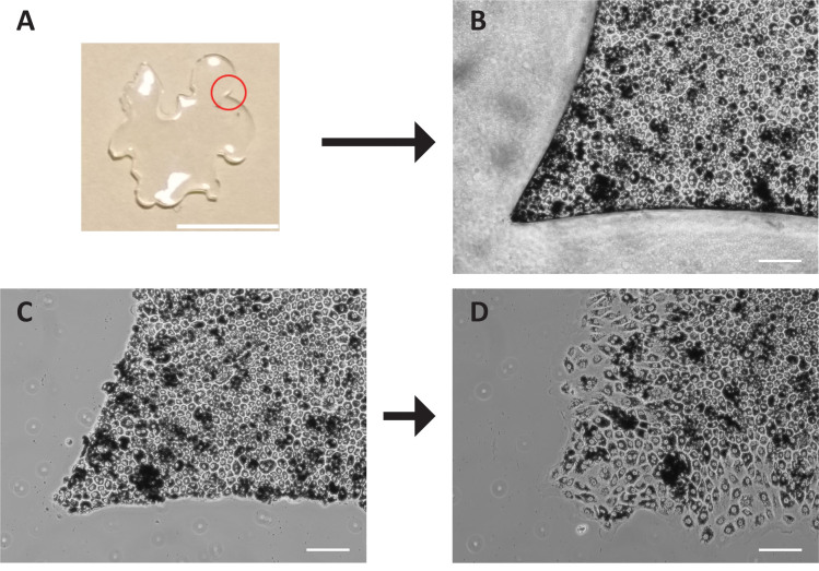
Methods: Micropatterning was used to control the spatial arrangement of primary porcine RPE cells using polydimethylsiloxane (PDMS) stencils. Patterns were formed into PDMS stencils to mimic 10%, 25%, and 50% overall detachment of the RPE monolayer. Zonula-occludens-1 (ZO-1), Ang-2, and VEGF were visualized using immunocytochemical (ICC) staining. An enzyme-linked immunosorbent assay (ELISA) was used to quantify extracellular Ang-2, VEGF, TIMP-1, and TIMP-2 levels. A rod outer segment (OS) phagocytosis assay was performed to determine how RPE junction loss directly affects photoreceptor support.
Results: The growth of primary porcine RPE cells was successfully controlled using stencils. Morphological changes and a decrease in pigmentation were observed, showing a decline in barrier and light absorption functions as degeneration increased. One day after stencil removal, junction proteins were delocalized, and angiogenic factor secretions were correlated with increased levels of detachment. Secretion levels of Ang-2 and TIMP-1 were significantly increased, whereas VEGF and TIMP-2 concentrations were not as affected by varying levels of detachment. OS phagocytosis appeared lower in RPE cells when ZO-1 was affected.
Conclusions: These results suggest a correlation between loss of junctions, abnormal angiogenic protein secretion, and reduced OS phagocytosis. Furthermore, Ang-2 and TIMP-1 proteins might be beneficial targets for AMD treatments, and their roles in retinal diseases deserve further investigation.
期刊介绍:
Molecular Vision is a peer-reviewed journal dedicated to the dissemination of research results in molecular biology, cell biology, and the genetics of the visual system (ocular and cortical).
Molecular Vision publishes articles presenting original research that has not previously been published and comprehensive articles reviewing the current status of a particular field or topic. Submissions to Molecular Vision are subjected to rigorous peer review. Molecular Vision does NOT publish preprints.
For authors, Molecular Vision provides a rapid means of communicating important results. Access to Molecular Vision is free and unrestricted, allowing the widest possible audience for your article. Digital publishing allows you to use color images freely (and without fees). Additionally, you may publish animations, sounds, or other supplementary information that clarifies or supports your article. Each of the authors of an article may also list an electronic mail address (which will be updated upon request) to give interested readers easy access to authors.
 分享
分享


 求助内容:
求助内容: 应助结果提醒方式:
 扫码关注我们
