Kana Mardhiyyah, Tanto Hariyanto, Teguh Wahju Sardjono, Sri Winarsih, Tatit Nurseta, Loeki Enggar Fitri
{"title":"胎儿生长迟缓与疟疾妊娠小鼠胎盘高凋亡细胞和低VEGF表达有关","authors":"Kana Mardhiyyah, Tanto Hariyanto, Teguh Wahju Sardjono, Sri Winarsih, Tatit Nurseta, Loeki Enggar Fitri","doi":"10.5455/medarh.2023.77.258-262","DOIUrl":null,"url":null,"abstract":"<p><strong>Background: </strong>During pregnancy, pregnant women are susceptible to malaria, contributing significantly to maternal and infant mortality.</p><p><strong>Objective: </strong>This research was conducted to study the effect of Plasmodium berghei infection in pregnant mice on fetal growth retardation through placental cell apoptosis and the change of local vascularization.</p><p><strong>Methods: </strong>Eighteen pregnant Balb/c strain mice resulting from simultanously mating were divided into two groups those were nine pregnant mice used as non infected group and nine pregnant mice infected with Plasmodium berghei on day 9th post mating used as infected group respectively. On day 15th of post mating, all of the pregnant mice were killed. Fetal weights were measured using analytic balance. Apoptosis of placental cells and VEGF expression in the placental tissue were measured using immunohistochemistry.</p><p><strong>Results: </strong>Result showed that there was sequestration of parasite-infected red blood cells (PRBCs) in intervillous space. Statistical analysis showed that the fetal weights in infected pregnant mice group was significantly lower than non infected one (p = 0.01), and the placental cell apoptosis in placental tissue of infected pregnant mice was significantly higher than the non infected one (p=0.00).There was also a significant difference on VEGF expression between infected group and non infected group (p= 0,00).</p><p><strong>Conclusion: </strong>Plasmodium berghei infection in pregnant Balb/c mice can cause fetal growth retardation due to high of placental cell apoptosis and low VEGF expression.</p>","PeriodicalId":94135,"journal":{"name":"Medical archives (Sarajevo, Bosnia and Herzegovina)","volume":"77 4","pages":"258-262"},"PeriodicalIF":0.0000,"publicationDate":"2023-01-01","publicationTypes":"Journal Article","fieldsOfStudy":null,"isOpenAccess":false,"openAccessPdf":"https://ftp.ncbi.nlm.nih.gov/pub/pmc/oa_pdf/aa/8d/medarch-77-258.PMC10591248.pdf","citationCount":"0","resultStr":"{\"title\":\"Fetal Growth Retardation is Associated with High Apoptotic Cells and Low VEGF Expression in Placenta of Malarial Pregnant Mice.\",\"authors\":\"Kana Mardhiyyah, Tanto Hariyanto, Teguh Wahju Sardjono, Sri Winarsih, Tatit Nurseta, Loeki Enggar Fitri\",\"doi\":\"10.5455/medarh.2023.77.258-262\",\"DOIUrl\":null,\"url\":null,\"abstract\":\"<p><strong>Background: </strong>During pregnancy, pregnant women are susceptible to malaria, contributing significantly to maternal and infant mortality.</p><p><strong>Objective: </strong>This research was conducted to study the effect of Plasmodium berghei infection in pregnant mice on fetal growth retardation through placental cell apoptosis and the change of local vascularization.</p><p><strong>Methods: </strong>Eighteen pregnant Balb/c strain mice resulting from simultanously mating were divided into two groups those were nine pregnant mice used as non infected group and nine pregnant mice infected with Plasmodium berghei on day 9th post mating used as infected group respectively. On day 15th of post mating, all of the pregnant mice were killed. Fetal weights were measured using analytic balance. Apoptosis of placental cells and VEGF expression in the placental tissue were measured using immunohistochemistry.</p><p><strong>Results: </strong>Result showed that there was sequestration of parasite-infected red blood cells (PRBCs) in intervillous space. Statistical analysis showed that the fetal weights in infected pregnant mice group was significantly lower than non infected one (p = 0.01), and the placental cell apoptosis in placental tissue of infected pregnant mice was significantly higher than the non infected one (p=0.00).There was also a significant difference on VEGF expression between infected group and non infected group (p= 0,00).</p><p><strong>Conclusion: </strong>Plasmodium berghei infection in pregnant Balb/c mice can cause fetal growth retardation due to high of placental cell apoptosis and low VEGF expression.</p>\",\"PeriodicalId\":94135,\"journal\":{\"name\":\"Medical archives (Sarajevo, Bosnia and Herzegovina)\",\"volume\":\"77 4\",\"pages\":\"258-262\"},\"PeriodicalIF\":0.0000,\"publicationDate\":\"2023-01-01\",\"publicationTypes\":\"Journal Article\",\"fieldsOfStudy\":null,\"isOpenAccess\":false,\"openAccessPdf\":\"https://ftp.ncbi.nlm.nih.gov/pub/pmc/oa_pdf/aa/8d/medarch-77-258.PMC10591248.pdf\",\"citationCount\":\"0\",\"resultStr\":null,\"platform\":\"Semanticscholar\",\"paperid\":null,\"PeriodicalName\":\"Medical archives (Sarajevo, Bosnia and Herzegovina)\",\"FirstCategoryId\":\"1085\",\"ListUrlMain\":\"https://doi.org/10.5455/medarh.2023.77.258-262\",\"RegionNum\":0,\"RegionCategory\":null,\"ArticlePicture\":[],\"TitleCN\":null,\"AbstractTextCN\":null,\"PMCID\":null,\"EPubDate\":\"\",\"PubModel\":\"\",\"JCR\":\"\",\"JCRName\":\"\",\"Score\":null,\"Total\":0}","platform":"Semanticscholar","paperid":null,"PeriodicalName":"Medical archives (Sarajevo, Bosnia and Herzegovina)","FirstCategoryId":"1085","ListUrlMain":"https://doi.org/10.5455/medarh.2023.77.258-262","RegionNum":0,"RegionCategory":null,"ArticlePicture":[],"TitleCN":null,"AbstractTextCN":null,"PMCID":null,"EPubDate":"","PubModel":"","JCR":"","JCRName":"","Score":null,"Total":0}
Fetal Growth Retardation is Associated with High Apoptotic Cells and Low VEGF Expression in Placenta of Malarial Pregnant Mice.
Background: During pregnancy, pregnant women are susceptible to malaria, contributing significantly to maternal and infant mortality.
Objective: This research was conducted to study the effect of Plasmodium berghei infection in pregnant mice on fetal growth retardation through placental cell apoptosis and the change of local vascularization.
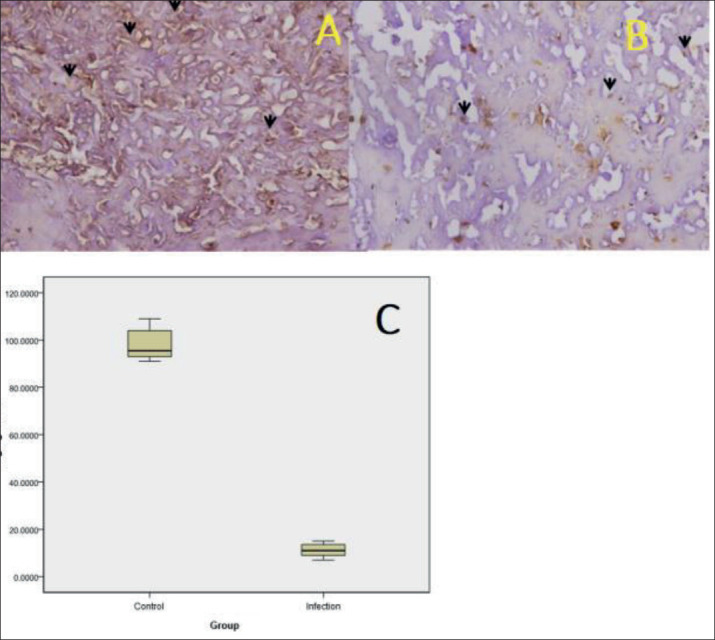
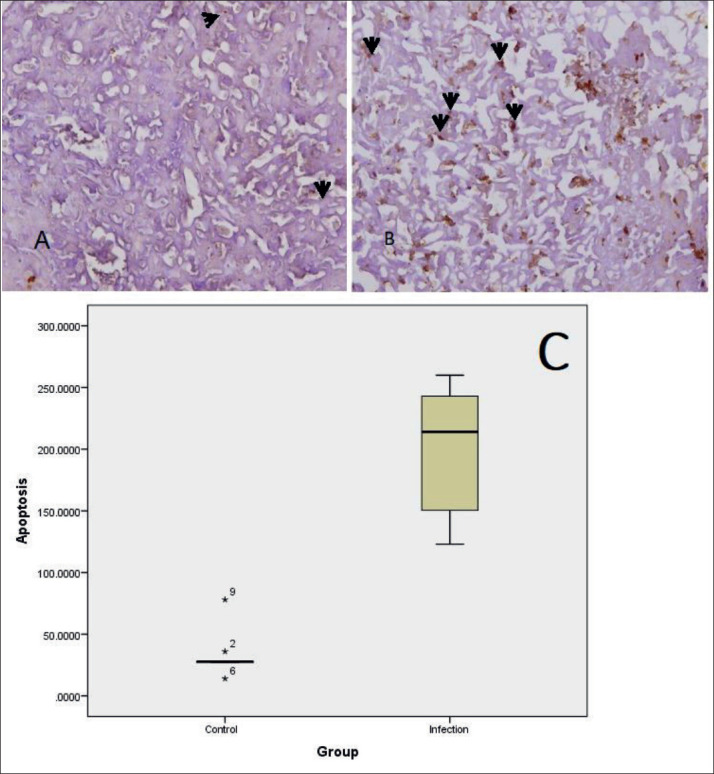
Methods: Eighteen pregnant Balb/c strain mice resulting from simultanously mating were divided into two groups those were nine pregnant mice used as non infected group and nine pregnant mice infected with Plasmodium berghei on day 9th post mating used as infected group respectively. On day 15th of post mating, all of the pregnant mice were killed. Fetal weights were measured using analytic balance. Apoptosis of placental cells and VEGF expression in the placental tissue were measured using immunohistochemistry.
Results: Result showed that there was sequestration of parasite-infected red blood cells (PRBCs) in intervillous space. Statistical analysis showed that the fetal weights in infected pregnant mice group was significantly lower than non infected one (p = 0.01), and the placental cell apoptosis in placental tissue of infected pregnant mice was significantly higher than the non infected one (p=0.00).There was also a significant difference on VEGF expression between infected group and non infected group (p= 0,00).
Conclusion: Plasmodium berghei infection in pregnant Balb/c mice can cause fetal growth retardation due to high of placental cell apoptosis and low VEGF expression.


 分享
分享

 求助内容:
求助内容: 应助结果提醒方式:
 扫码关注我们
