{"title":"传统二维磁共振成像诊断腰骶部椎管外狭窄的可靠性","authors":"Kohei Takahashi, Myo Min Latt, Takumi Tsubakino, Manabu Suzuki, Takeshi Nakamura, Takeshi Hoshikawa, Tomowaki Nakagawa, Ko Hashimoto, Takahiro Onoki, Toshimi Aizawa, Yasuhisa Tanaka","doi":"10.22603/ssrr.2023-0110","DOIUrl":null,"url":null,"abstract":"<p><strong>Introduction: </strong>Three-dimensional (3D) magnetic resonance imaging (MRI) is reportedly superior to two-dimensional (2D) MRI for diagnosing lumbar foraminal stenosis at L5-S1. In this study, we strictly distinguished the intra- and extraforaminal regions and compared the diagnostic reliability and accuracy of 2D and 3D MRI in each region.</p><p><strong>Methods: </strong>A total of 92 surgical cases of unilateral L5 radiculopathy were selected for imaging analysis, including 46 of foraminal stenosis at L5-S1 (Group F) and 46 of intraspinal canal stenosis at L4-5 (Group C) (48 men, 44 women; mean age, 66 years). The 2D and 3D MRI sets were assessed twice by two examiners. They were informed only of the laterality of the lesion in each case and asked to select among the following for each modality: \"absence of foraminal stenosis,\" \"intraforaminal stenosis,\" \"extraforaminal stenosis,\" and \"coincident intraforaminal and extraforaminal stenosis.\" The intra- and interobserver reliabilities were evaluated using kappa (κ) statistics for the intra- and extraforaminal regions and compared between 2D and 3D MRI. For each case, disagreements between examiners were resolved through discussion to obtain a diagnostic judgment for each modality. Subsequently, the final diagnosis of intra- and/or extraforaminal stenosis in Group F was made using multiple modalities and intraoperative findings. A comparison between 2D and 3D MRI in terms of diagnostic accuracy was performed for the intra- and extraforaminal regions.</p><p><strong>Results: </strong>No significant difference was observed in the κ statistics between 2D and 3D MRI for the intraforaminal region, whereas 3D MRI had significantly larger κ statistic than 2D MRI for the extraforaminal region. Ultimately, 3D MRI perfectly judged the extraforaminal region, whereas 2D MRI detected only 44.8% of the cases of extraforaminal stenosis.</p><p><strong>Conclusions: </strong>More than half of extraforaminal stenosis was overlooked by 2D MRI, suggesting that it is unreliable for diagnosing extraforaminal stenosis at L5-S1.</p>","PeriodicalId":22253,"journal":{"name":"Spine Surgery and Related Research","volume":"1 1","pages":"526-532"},"PeriodicalIF":1.6000,"publicationDate":"2023-08-10","publicationTypes":"Journal Article","fieldsOfStudy":null,"isOpenAccess":false,"openAccessPdf":"https://www.ncbi.nlm.nih.gov/pmc/articles/PMC10710884/pdf/","citationCount":"1","resultStr":"{\"title\":\"Reliability of Conventional Two-Dimensional Magnetic Resonance Imaging for Diagnosing Extraforaminal Stenosis in Lumbosacral Transition.\",\"authors\":\"Kohei Takahashi, Myo Min Latt, Takumi Tsubakino, Manabu Suzuki, Takeshi Nakamura, Takeshi Hoshikawa, Tomowaki Nakagawa, Ko Hashimoto, Takahiro Onoki, Toshimi Aizawa, Yasuhisa Tanaka\",\"doi\":\"10.22603/ssrr.2023-0110\",\"DOIUrl\":null,\"url\":null,\"abstract\":\"<p><strong>Introduction: </strong>Three-dimensional (3D) magnetic resonance imaging (MRI) is reportedly superior to two-dimensional (2D) MRI for diagnosing lumbar foraminal stenosis at L5-S1. In this study, we strictly distinguished the intra- and extraforaminal regions and compared the diagnostic reliability and accuracy of 2D and 3D MRI in each region.</p><p><strong>Methods: </strong>A total of 92 surgical cases of unilateral L5 radiculopathy were selected for imaging analysis, including 46 of foraminal stenosis at L5-S1 (Group F) and 46 of intraspinal canal stenosis at L4-5 (Group C) (48 men, 44 women; mean age, 66 years). The 2D and 3D MRI sets were assessed twice by two examiners. They were informed only of the laterality of the lesion in each case and asked to select among the following for each modality: \\\"absence of foraminal stenosis,\\\" \\\"intraforaminal stenosis,\\\" \\\"extraforaminal stenosis,\\\" and \\\"coincident intraforaminal and extraforaminal stenosis.\\\" The intra- and interobserver reliabilities were evaluated using kappa (κ) statistics for the intra- and extraforaminal regions and compared between 2D and 3D MRI. For each case, disagreements between examiners were resolved through discussion to obtain a diagnostic judgment for each modality. Subsequently, the final diagnosis of intra- and/or extraforaminal stenosis in Group F was made using multiple modalities and intraoperative findings. A comparison between 2D and 3D MRI in terms of diagnostic accuracy was performed for the intra- and extraforaminal regions.</p><p><strong>Results: </strong>No significant difference was observed in the κ statistics between 2D and 3D MRI for the intraforaminal region, whereas 3D MRI had significantly larger κ statistic than 2D MRI for the extraforaminal region. Ultimately, 3D MRI perfectly judged the extraforaminal region, whereas 2D MRI detected only 44.8% of the cases of extraforaminal stenosis.</p><p><strong>Conclusions: </strong>More than half of extraforaminal stenosis was overlooked by 2D MRI, suggesting that it is unreliable for diagnosing extraforaminal stenosis at L5-S1.</p>\",\"PeriodicalId\":22253,\"journal\":{\"name\":\"Spine Surgery and Related Research\",\"volume\":\"1 1\",\"pages\":\"526-532\"},\"PeriodicalIF\":1.6000,\"publicationDate\":\"2023-08-10\",\"publicationTypes\":\"Journal Article\",\"fieldsOfStudy\":null,\"isOpenAccess\":false,\"openAccessPdf\":\"https://www.ncbi.nlm.nih.gov/pmc/articles/PMC10710884/pdf/\",\"citationCount\":\"1\",\"resultStr\":null,\"platform\":\"Semanticscholar\",\"paperid\":null,\"PeriodicalName\":\"Spine Surgery and Related Research\",\"FirstCategoryId\":\"1085\",\"ListUrlMain\":\"https://doi.org/10.22603/ssrr.2023-0110\",\"RegionNum\":0,\"RegionCategory\":null,\"ArticlePicture\":[],\"TitleCN\":null,\"AbstractTextCN\":null,\"PMCID\":null,\"EPubDate\":\"2023/11/27 0:00:00\",\"PubModel\":\"eCollection\",\"JCR\":\"Q3\",\"JCRName\":\"SURGERY\",\"Score\":null,\"Total\":0}","platform":"Semanticscholar","paperid":null,"PeriodicalName":"Spine Surgery and Related Research","FirstCategoryId":"1085","ListUrlMain":"https://doi.org/10.22603/ssrr.2023-0110","RegionNum":0,"RegionCategory":null,"ArticlePicture":[],"TitleCN":null,"AbstractTextCN":null,"PMCID":null,"EPubDate":"2023/11/27 0:00:00","PubModel":"eCollection","JCR":"Q3","JCRName":"SURGERY","Score":null,"Total":0}
Reliability of Conventional Two-Dimensional Magnetic Resonance Imaging for Diagnosing Extraforaminal Stenosis in Lumbosacral Transition.
Introduction: Three-dimensional (3D) magnetic resonance imaging (MRI) is reportedly superior to two-dimensional (2D) MRI for diagnosing lumbar foraminal stenosis at L5-S1. In this study, we strictly distinguished the intra- and extraforaminal regions and compared the diagnostic reliability and accuracy of 2D and 3D MRI in each region.
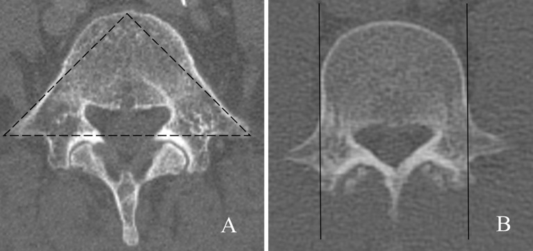
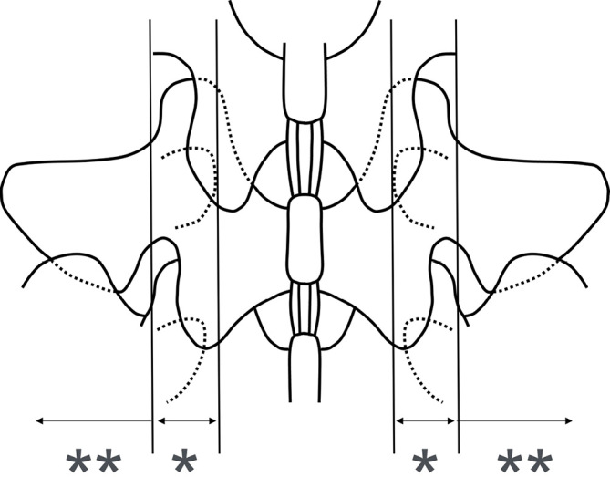
Methods: A total of 92 surgical cases of unilateral L5 radiculopathy were selected for imaging analysis, including 46 of foraminal stenosis at L5-S1 (Group F) and 46 of intraspinal canal stenosis at L4-5 (Group C) (48 men, 44 women; mean age, 66 years). The 2D and 3D MRI sets were assessed twice by two examiners. They were informed only of the laterality of the lesion in each case and asked to select among the following for each modality: "absence of foraminal stenosis," "intraforaminal stenosis," "extraforaminal stenosis," and "coincident intraforaminal and extraforaminal stenosis." The intra- and interobserver reliabilities were evaluated using kappa (κ) statistics for the intra- and extraforaminal regions and compared between 2D and 3D MRI. For each case, disagreements between examiners were resolved through discussion to obtain a diagnostic judgment for each modality. Subsequently, the final diagnosis of intra- and/or extraforaminal stenosis in Group F was made using multiple modalities and intraoperative findings. A comparison between 2D and 3D MRI in terms of diagnostic accuracy was performed for the intra- and extraforaminal regions.
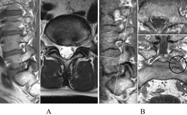
Results: No significant difference was observed in the κ statistics between 2D and 3D MRI for the intraforaminal region, whereas 3D MRI had significantly larger κ statistic than 2D MRI for the extraforaminal region. Ultimately, 3D MRI perfectly judged the extraforaminal region, whereas 2D MRI detected only 44.8% of the cases of extraforaminal stenosis.
Conclusions: More than half of extraforaminal stenosis was overlooked by 2D MRI, suggesting that it is unreliable for diagnosing extraforaminal stenosis at L5-S1.


 分享
分享
 求助内容:
求助内容: 应助结果提醒方式:
 扫码关注我们
