Rodolfo Dias Chiari-Correia, Vitor Tumas, Antônio Carlos Santos, Carlos Ernesto G Salmon
{"title":"伴有抑郁症状和不伴有抑郁症状的 MCI 患者大脑结构和功能差异及其与阿尔茨海默病的关系:磁共振成像研究。","authors":"Rodolfo Dias Chiari-Correia, Vitor Tumas, Antônio Carlos Santos, Carlos Ernesto G Salmon","doi":"10.1093/psyrad/kkad008","DOIUrl":null,"url":null,"abstract":"<p><strong>Background: </strong>The mild cognitive impairment (MCI) stage among elderly individuals is very complex, and the level of diagnostic accuracy is far from ideal. Some studies have tried to improve the 'MCI due to Alzheimer's disease (AD)' classification by further stratifying these patients into subgroups. Depression-related symptoms may play an important role in helping to better define the MCI stage in elderly individuals.</p><p><strong>Objective: </strong>In this work, we explored functional and structural differences in the brains of patients with nondepressed MCI (nDMCI) and patients with MCI with depressive symptoms (DMCI), and we examined how these groups relate to AD atrophy patterns and cognitive functioning.</p><p><strong>Methods: </strong>Sixty-five participants underwent MRI exams and were divided into four groups: cognitively normal, nDMCI, DMCI, and AD. We compared the regional brain volumes, cortical thickness, and white matter microstructure measures using diffusion tensor imaging among groups. Additionally, we evaluated changes in functional connectivity using fMRI data.</p><p><strong>Results: </strong>In comparison to the nDMCI group, the DMCI patients had more pronounced atrophy in the hippocampus and amygdala. Additionally, DMCI patients had asymmetric damage in the limbic-frontal white matter connection. Furthermore, two medial posterior regions, the isthmus of cingulate gyrus and especially the lingual gyrus, had high importance in the structural and functional differentiation between the two groups.</p><p><strong>Conclusion: </strong>It is possible to differentiate nDMCI from DMCI patients using MRI techniques, which may contribute to a better characterization of subtypes of the MCI stage.</p>","PeriodicalId":93496,"journal":{"name":"Psychoradiology","volume":"1 1","pages":"kkad008"},"PeriodicalIF":2.9000,"publicationDate":"2023-06-13","publicationTypes":"Journal Article","fieldsOfStudy":null,"isOpenAccess":false,"openAccessPdf":"https://www.ncbi.nlm.nih.gov/pmc/articles/PMC10917365/pdf/","citationCount":"0","resultStr":"{\"title\":\"Structural and functional differences in the brains of patients with MCI with and without depressive symptoms and their relations with Alzheimer's disease: an MRI study.\",\"authors\":\"Rodolfo Dias Chiari-Correia, Vitor Tumas, Antônio Carlos Santos, Carlos Ernesto G Salmon\",\"doi\":\"10.1093/psyrad/kkad008\",\"DOIUrl\":null,\"url\":null,\"abstract\":\"<p><strong>Background: </strong>The mild cognitive impairment (MCI) stage among elderly individuals is very complex, and the level of diagnostic accuracy is far from ideal. Some studies have tried to improve the 'MCI due to Alzheimer's disease (AD)' classification by further stratifying these patients into subgroups. Depression-related symptoms may play an important role in helping to better define the MCI stage in elderly individuals.</p><p><strong>Objective: </strong>In this work, we explored functional and structural differences in the brains of patients with nondepressed MCI (nDMCI) and patients with MCI with depressive symptoms (DMCI), and we examined how these groups relate to AD atrophy patterns and cognitive functioning.</p><p><strong>Methods: </strong>Sixty-five participants underwent MRI exams and were divided into four groups: cognitively normal, nDMCI, DMCI, and AD. We compared the regional brain volumes, cortical thickness, and white matter microstructure measures using diffusion tensor imaging among groups. Additionally, we evaluated changes in functional connectivity using fMRI data.</p><p><strong>Results: </strong>In comparison to the nDMCI group, the DMCI patients had more pronounced atrophy in the hippocampus and amygdala. Additionally, DMCI patients had asymmetric damage in the limbic-frontal white matter connection. Furthermore, two medial posterior regions, the isthmus of cingulate gyrus and especially the lingual gyrus, had high importance in the structural and functional differentiation between the two groups.</p><p><strong>Conclusion: </strong>It is possible to differentiate nDMCI from DMCI patients using MRI techniques, which may contribute to a better characterization of subtypes of the MCI stage.</p>\",\"PeriodicalId\":93496,\"journal\":{\"name\":\"Psychoradiology\",\"volume\":\"1 1\",\"pages\":\"kkad008\"},\"PeriodicalIF\":2.9000,\"publicationDate\":\"2023-06-13\",\"publicationTypes\":\"Journal Article\",\"fieldsOfStudy\":null,\"isOpenAccess\":false,\"openAccessPdf\":\"https://www.ncbi.nlm.nih.gov/pmc/articles/PMC10917365/pdf/\",\"citationCount\":\"0\",\"resultStr\":null,\"platform\":\"Semanticscholar\",\"paperid\":null,\"PeriodicalName\":\"Psychoradiology\",\"FirstCategoryId\":\"1085\",\"ListUrlMain\":\"https://doi.org/10.1093/psyrad/kkad008\",\"RegionNum\":0,\"RegionCategory\":null,\"ArticlePicture\":[],\"TitleCN\":null,\"AbstractTextCN\":null,\"PMCID\":null,\"EPubDate\":\"2023/1/1 0:00:00\",\"PubModel\":\"eCollection\",\"JCR\":\"\",\"JCRName\":\"\",\"Score\":null,\"Total\":0}","platform":"Semanticscholar","paperid":null,"PeriodicalName":"Psychoradiology","FirstCategoryId":"1085","ListUrlMain":"https://doi.org/10.1093/psyrad/kkad008","RegionNum":0,"RegionCategory":null,"ArticlePicture":[],"TitleCN":null,"AbstractTextCN":null,"PMCID":null,"EPubDate":"2023/1/1 0:00:00","PubModel":"eCollection","JCR":"","JCRName":"","Score":null,"Total":0}
Structural and functional differences in the brains of patients with MCI with and without depressive symptoms and their relations with Alzheimer's disease: an MRI study.
Background: The mild cognitive impairment (MCI) stage among elderly individuals is very complex, and the level of diagnostic accuracy is far from ideal. Some studies have tried to improve the 'MCI due to Alzheimer's disease (AD)' classification by further stratifying these patients into subgroups. Depression-related symptoms may play an important role in helping to better define the MCI stage in elderly individuals.
Objective: In this work, we explored functional and structural differences in the brains of patients with nondepressed MCI (nDMCI) and patients with MCI with depressive symptoms (DMCI), and we examined how these groups relate to AD atrophy patterns and cognitive functioning.
Methods: Sixty-five participants underwent MRI exams and were divided into four groups: cognitively normal, nDMCI, DMCI, and AD. We compared the regional brain volumes, cortical thickness, and white matter microstructure measures using diffusion tensor imaging among groups. Additionally, we evaluated changes in functional connectivity using fMRI data.
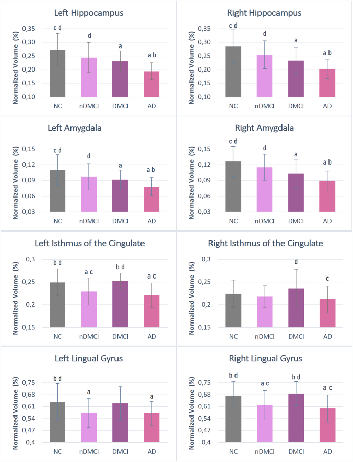
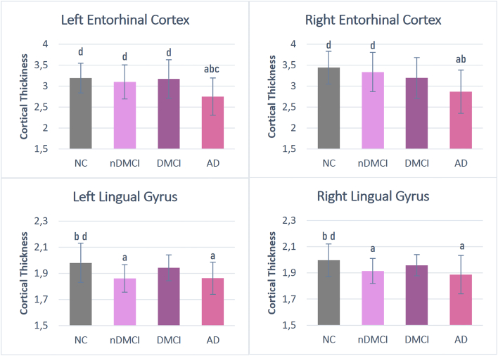
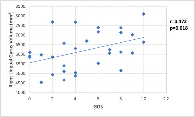
Results: In comparison to the nDMCI group, the DMCI patients had more pronounced atrophy in the hippocampus and amygdala. Additionally, DMCI patients had asymmetric damage in the limbic-frontal white matter connection. Furthermore, two medial posterior regions, the isthmus of cingulate gyrus and especially the lingual gyrus, had high importance in the structural and functional differentiation between the two groups.
Conclusion: It is possible to differentiate nDMCI from DMCI patients using MRI techniques, which may contribute to a better characterization of subtypes of the MCI stage.


 分享
分享

 求助内容:
求助内容: 应助结果提醒方式:
 扫码关注我们
