{"title":"shoji框架刺穿眶颅伤:罕见的日式住宅室内事故。","authors":"Hideaki Ueno, Satoshi Tsutsumi, Yasutoshi Akasaki, Kohei Yoshida, Natsuki Sugiyama, Hisato Ishii","doi":"10.25259/SNI_29_2023","DOIUrl":null,"url":null,"abstract":"<p><strong>Background: </strong>To the best of our knowledge, there are no reports of penetrating orbitocranial injury (POCI) caused by a shoji frame.</p><p><strong>Case description: </strong>A 68-year-old man fell in his living room and was stuck headfirst by a shoji frame. At presentation, marked swelling was noted in the right upper eyelid, with the edge of the broken shoji frame exposed superficially. Computed tomography (CT) revealed a hypodense linear structure located in the upper lateral sector of the orbit, partially protruding into the middle cranial fossa. Contrast-enhanced CT revealed intact ophthalmic artery and superior ophthalmic vein. The patient was managed with frontotemporal craniotomy. The shoji frame was extracted by pushing out the extradurally located proximal edge from the cranial cavity and simultaneously pulling the distal edge from the stab wound in the upper eyelid. Postoperatively, the patient received intravenous antibiotic therapy for 18 days.</p><p><strong>Conclusion: </strong>POCI can be caused by shoji frames as a result of an indoor accident. The broken shoji frame is evidently delineated on CT, which can result in prompt extraction.</p>","PeriodicalId":38981,"journal":{"name":"Surgical Neurology International","volume":"14 ","pages":"51"},"PeriodicalIF":0.0000,"publicationDate":"2023-01-01","publicationTypes":"Journal Article","fieldsOfStudy":null,"isOpenAccess":false,"openAccessPdf":"https://ftp.ncbi.nlm.nih.gov/pub/pmc/oa_pdf/8e/d4/SNI-14-51.PMC9990803.pdf","citationCount":"0","resultStr":"{\"title\":\"Penetrating orbitocranial injury by shoji frame: A rare indoor accident in a Japanese style house.\",\"authors\":\"Hideaki Ueno, Satoshi Tsutsumi, Yasutoshi Akasaki, Kohei Yoshida, Natsuki Sugiyama, Hisato Ishii\",\"doi\":\"10.25259/SNI_29_2023\",\"DOIUrl\":null,\"url\":null,\"abstract\":\"<p><strong>Background: </strong>To the best of our knowledge, there are no reports of penetrating orbitocranial injury (POCI) caused by a shoji frame.</p><p><strong>Case description: </strong>A 68-year-old man fell in his living room and was stuck headfirst by a shoji frame. At presentation, marked swelling was noted in the right upper eyelid, with the edge of the broken shoji frame exposed superficially. Computed tomography (CT) revealed a hypodense linear structure located in the upper lateral sector of the orbit, partially protruding into the middle cranial fossa. Contrast-enhanced CT revealed intact ophthalmic artery and superior ophthalmic vein. The patient was managed with frontotemporal craniotomy. The shoji frame was extracted by pushing out the extradurally located proximal edge from the cranial cavity and simultaneously pulling the distal edge from the stab wound in the upper eyelid. Postoperatively, the patient received intravenous antibiotic therapy for 18 days.</p><p><strong>Conclusion: </strong>POCI can be caused by shoji frames as a result of an indoor accident. The broken shoji frame is evidently delineated on CT, which can result in prompt extraction.</p>\",\"PeriodicalId\":38981,\"journal\":{\"name\":\"Surgical Neurology International\",\"volume\":\"14 \",\"pages\":\"51\"},\"PeriodicalIF\":0.0000,\"publicationDate\":\"2023-01-01\",\"publicationTypes\":\"Journal Article\",\"fieldsOfStudy\":null,\"isOpenAccess\":false,\"openAccessPdf\":\"https://ftp.ncbi.nlm.nih.gov/pub/pmc/oa_pdf/8e/d4/SNI-14-51.PMC9990803.pdf\",\"citationCount\":\"0\",\"resultStr\":null,\"platform\":\"Semanticscholar\",\"paperid\":null,\"PeriodicalName\":\"Surgical Neurology International\",\"FirstCategoryId\":\"1085\",\"ListUrlMain\":\"https://doi.org/10.25259/SNI_29_2023\",\"RegionNum\":0,\"RegionCategory\":null,\"ArticlePicture\":[],\"TitleCN\":null,\"AbstractTextCN\":null,\"PMCID\":null,\"EPubDate\":\"\",\"PubModel\":\"\",\"JCR\":\"Q3\",\"JCRName\":\"Medicine\",\"Score\":null,\"Total\":0}","platform":"Semanticscholar","paperid":null,"PeriodicalName":"Surgical Neurology International","FirstCategoryId":"1085","ListUrlMain":"https://doi.org/10.25259/SNI_29_2023","RegionNum":0,"RegionCategory":null,"ArticlePicture":[],"TitleCN":null,"AbstractTextCN":null,"PMCID":null,"EPubDate":"","PubModel":"","JCR":"Q3","JCRName":"Medicine","Score":null,"Total":0}
Penetrating orbitocranial injury by shoji frame: A rare indoor accident in a Japanese style house.
Background: To the best of our knowledge, there are no reports of penetrating orbitocranial injury (POCI) caused by a shoji frame.
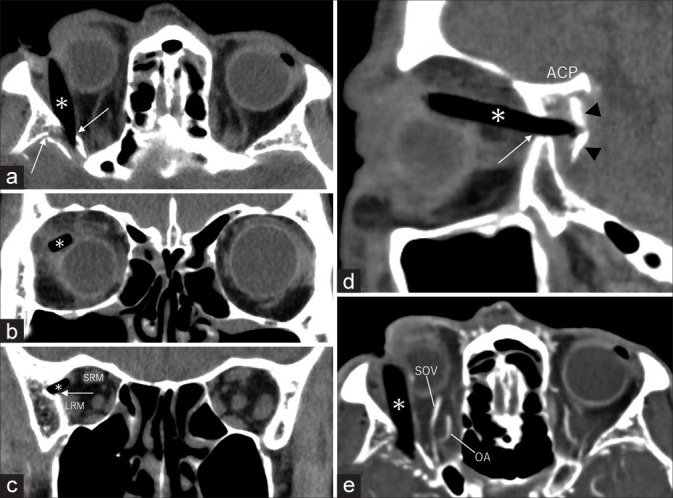
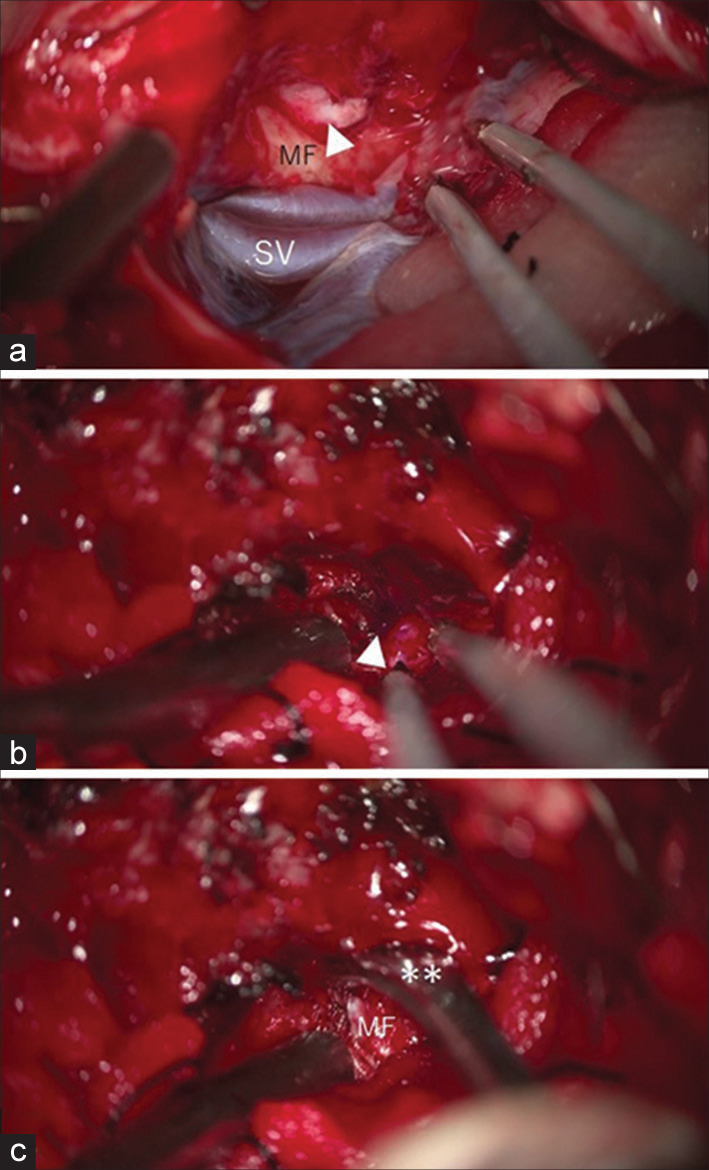
Case description: A 68-year-old man fell in his living room and was stuck headfirst by a shoji frame. At presentation, marked swelling was noted in the right upper eyelid, with the edge of the broken shoji frame exposed superficially. Computed tomography (CT) revealed a hypodense linear structure located in the upper lateral sector of the orbit, partially protruding into the middle cranial fossa. Contrast-enhanced CT revealed intact ophthalmic artery and superior ophthalmic vein. The patient was managed with frontotemporal craniotomy. The shoji frame was extracted by pushing out the extradurally located proximal edge from the cranial cavity and simultaneously pulling the distal edge from the stab wound in the upper eyelid. Postoperatively, the patient received intravenous antibiotic therapy for 18 days.
Conclusion: POCI can be caused by shoji frames as a result of an indoor accident. The broken shoji frame is evidently delineated on CT, which can result in prompt extraction.


 分享
分享
 求助内容:
求助内容: 应助结果提醒方式:
 扫码关注我们
