Campbell Goldsmith, Jennifer Cheng, Douglas Mintz, Peter Moley
{"title":"股骨版本测量与股骨髋臼撞击相关主诉患者计算机断层扫描和磁共振成像研究之间的相关性","authors":"Campbell Goldsmith, Jennifer Cheng, Douglas Mintz, Peter Moley","doi":"10.1093/jhps/hnac036","DOIUrl":null,"url":null,"abstract":"<p><p>Computed tomography (CT) is considered the gold standard for femoral version measurement. However, recent data have shown magnetic resonance imaging (MRI) as another modality to measure femoral version. This study aimed to correlate MRI and CT femoral version measurements in patients presenting with a femoroacetabular impingement (FAI)-related complaint. Patients (18-35 years old) who presented to the hip preservation clinic and radiology department with a suspected FAI diagnosis from 26 December 2018 to 4 March 2020 were included. All patients had a CT and MRI of the hip, with images including both hips and knees, as per our institution's protocol for possible hip preservation surgery. Patients were excluded if they were missing views of the knees, or if they had a history or imaging appearance of any condition affecting femoral version at the femoral head (e.g. slipped capital femoral epiphysis). Femoral version was measured by three reviewers. Fifty-eight patients were included, and 36 (62%) were female. Femoral version averaged 6.1° ± 11.8° on CT and 6.5° ± 10.8° on MRI. A strong positive correlation was reported between the two imaging modalities (<i>r</i>: 0.81; <i>P</i> < 0.001). Inter-rater reliability among the three reviewers was excellent and statistically significant for measurements on both MRI [intraclass correlation coefficient (ICC): 0.95; 95% CI: 0.85, 0.99; <i>P</i> < 0.001] and CT (ICC: 0.97; 95% CI: 0.92, 0.99; <i>P</i> < 0.001). Our finding suggests that MRI is a sufficient method for measuring femoral version to determine disease etiology and treatment progression. To avoid exposing patients to ionizing radiation, physicians should not obtain CT scans to evaluate femoral version.</p>","PeriodicalId":48583,"journal":{"name":"Journal of Hip Preservation Surgery","volume":"9 4","pages":"219-224"},"PeriodicalIF":1.1000,"publicationDate":"2022-12-01","publicationTypes":"Journal Article","fieldsOfStudy":null,"isOpenAccess":false,"openAccessPdf":"https://ftp.ncbi.nlm.nih.gov/pub/pmc/oa_pdf/b1/1f/hnac036.PMC9993453.pdf","citationCount":"2","resultStr":"{\"title\":\"Correlation of femoral version measurements between computed tomography and magnetic resonance imaging studies in patients presenting with a femoroacetabular impingement-related complaint.\",\"authors\":\"Campbell Goldsmith, Jennifer Cheng, Douglas Mintz, Peter Moley\",\"doi\":\"10.1093/jhps/hnac036\",\"DOIUrl\":null,\"url\":null,\"abstract\":\"<p><p>Computed tomography (CT) is considered the gold standard for femoral version measurement. However, recent data have shown magnetic resonance imaging (MRI) as another modality to measure femoral version. This study aimed to correlate MRI and CT femoral version measurements in patients presenting with a femoroacetabular impingement (FAI)-related complaint. Patients (18-35 years old) who presented to the hip preservation clinic and radiology department with a suspected FAI diagnosis from 26 December 2018 to 4 March 2020 were included. All patients had a CT and MRI of the hip, with images including both hips and knees, as per our institution's protocol for possible hip preservation surgery. Patients were excluded if they were missing views of the knees, or if they had a history or imaging appearance of any condition affecting femoral version at the femoral head (e.g. slipped capital femoral epiphysis). Femoral version was measured by three reviewers. Fifty-eight patients were included, and 36 (62%) were female. Femoral version averaged 6.1° ± 11.8° on CT and 6.5° ± 10.8° on MRI. A strong positive correlation was reported between the two imaging modalities (<i>r</i>: 0.81; <i>P</i> < 0.001). Inter-rater reliability among the three reviewers was excellent and statistically significant for measurements on both MRI [intraclass correlation coefficient (ICC): 0.95; 95% CI: 0.85, 0.99; <i>P</i> < 0.001] and CT (ICC: 0.97; 95% CI: 0.92, 0.99; <i>P</i> < 0.001). Our finding suggests that MRI is a sufficient method for measuring femoral version to determine disease etiology and treatment progression. To avoid exposing patients to ionizing radiation, physicians should not obtain CT scans to evaluate femoral version.</p>\",\"PeriodicalId\":48583,\"journal\":{\"name\":\"Journal of Hip Preservation Surgery\",\"volume\":\"9 4\",\"pages\":\"219-224\"},\"PeriodicalIF\":1.1000,\"publicationDate\":\"2022-12-01\",\"publicationTypes\":\"Journal Article\",\"fieldsOfStudy\":null,\"isOpenAccess\":false,\"openAccessPdf\":\"https://ftp.ncbi.nlm.nih.gov/pub/pmc/oa_pdf/b1/1f/hnac036.PMC9993453.pdf\",\"citationCount\":\"2\",\"resultStr\":null,\"platform\":\"Semanticscholar\",\"paperid\":null,\"PeriodicalName\":\"Journal of Hip Preservation Surgery\",\"FirstCategoryId\":\"3\",\"ListUrlMain\":\"https://doi.org/10.1093/jhps/hnac036\",\"RegionNum\":4,\"RegionCategory\":\"医学\",\"ArticlePicture\":[],\"TitleCN\":null,\"AbstractTextCN\":null,\"PMCID\":null,\"EPubDate\":\"\",\"PubModel\":\"\",\"JCR\":\"Q3\",\"JCRName\":\"ORTHOPEDICS\",\"Score\":null,\"Total\":0}","platform":"Semanticscholar","paperid":null,"PeriodicalName":"Journal of Hip Preservation Surgery","FirstCategoryId":"3","ListUrlMain":"https://doi.org/10.1093/jhps/hnac036","RegionNum":4,"RegionCategory":"医学","ArticlePicture":[],"TitleCN":null,"AbstractTextCN":null,"PMCID":null,"EPubDate":"","PubModel":"","JCR":"Q3","JCRName":"ORTHOPEDICS","Score":null,"Total":0}
引用次数: 2
摘要
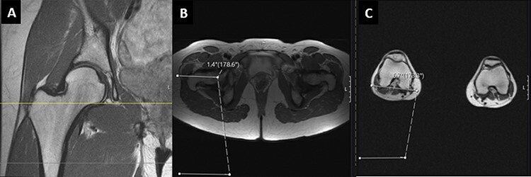
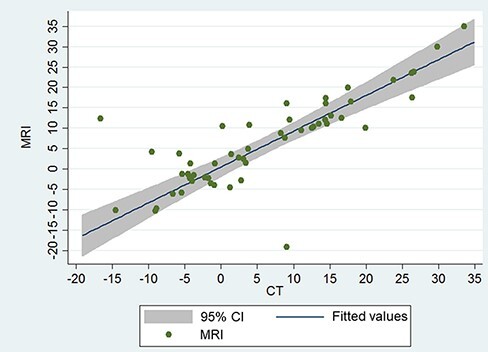
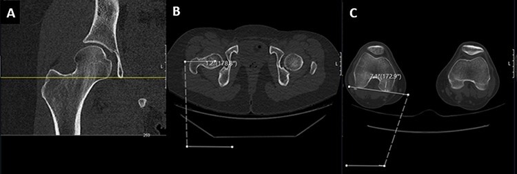
计算机断层扫描(CT)被认为是股骨版本测量的金标准。然而,最近的数据显示磁共振成像(MRI)是另一种测量股骨版本的方式。本研究旨在将股骨髋臼撞击(FAI)相关主诉患者的MRI和CT股骨版本测量相关联。纳入了2018年12月26日至2020年3月4日期间髋关节保存诊所和放射科疑似FAI诊断的患者(18-35岁)。所有患者都进行了髋关节的CT和MRI检查,包括髋关节和膝关节的图像,按照我们机构可能的髋关节保留手术的协议。如果患者缺少膝关节视图,或者如果他们有任何影响股骨头股骨变形的病史或影像学表现(如股骨头骨骺滑动),则排除在外。股骨版本由三位评论者测量。纳入58例患者,其中36例(62%)为女性。股骨型CT平均6.1°±11.8°,MRI平均6.5°±10.8°。两种成像方式之间有很强的正相关(r: 0.81;p p p
Correlation of femoral version measurements between computed tomography and magnetic resonance imaging studies in patients presenting with a femoroacetabular impingement-related complaint.
Computed tomography (CT) is considered the gold standard for femoral version measurement. However, recent data have shown magnetic resonance imaging (MRI) as another modality to measure femoral version. This study aimed to correlate MRI and CT femoral version measurements in patients presenting with a femoroacetabular impingement (FAI)-related complaint. Patients (18-35 years old) who presented to the hip preservation clinic and radiology department with a suspected FAI diagnosis from 26 December 2018 to 4 March 2020 were included. All patients had a CT and MRI of the hip, with images including both hips and knees, as per our institution's protocol for possible hip preservation surgery. Patients were excluded if they were missing views of the knees, or if they had a history or imaging appearance of any condition affecting femoral version at the femoral head (e.g. slipped capital femoral epiphysis). Femoral version was measured by three reviewers. Fifty-eight patients were included, and 36 (62%) were female. Femoral version averaged 6.1° ± 11.8° on CT and 6.5° ± 10.8° on MRI. A strong positive correlation was reported between the two imaging modalities (r: 0.81; P < 0.001). Inter-rater reliability among the three reviewers was excellent and statistically significant for measurements on both MRI [intraclass correlation coefficient (ICC): 0.95; 95% CI: 0.85, 0.99; P < 0.001] and CT (ICC: 0.97; 95% CI: 0.92, 0.99; P < 0.001). Our finding suggests that MRI is a sufficient method for measuring femoral version to determine disease etiology and treatment progression. To avoid exposing patients to ionizing radiation, physicians should not obtain CT scans to evaluate femoral version.


 分享
分享
 求助内容:
求助内容: 应助结果提醒方式:
 扫码关注我们
