Dae Hwan Park, June Key Lee, Bong Soo Baik, Wan Suk Yang, Sun Young Kim
{"title":"7岁男孩创伤性颞浅动脉血肿性假性动脉瘤1例报告。","authors":"Dae Hwan Park, June Key Lee, Bong Soo Baik, Wan Suk Yang, Sun Young Kim","doi":"10.7181/acfs.2022.00703","DOIUrl":null,"url":null,"abstract":"<p><p>The superficial temporal artery (STA), the terminal branch of the external carotid artery, is divided into the frontal (anterior) and parietal (posterior) branches. The frontal branch of the STA is located superficially on the anterior region of the scalp, making it especially susceptible to trauma. Here, we report a traumatic pseudoaneurysm of the STA in a 7-year-old boy who was injured in a minor car accident. A physical examination showed only a small bruise on the patient's forehead, and all vital signs were stable at the emergency room of our medical center. A facial computed tomography scan showed no significant findings. However, the boy later re-visited the hospital with slight swelling on the right forehead, and an ultrasonography scan revealed a hematoma near the right temporal artery. The resected hematoma (approximately 2 cm) was diagnosed as a traumatic pseudoaneurysm. Awareness of the possibility of a traumatic pseudoaneurysm in the STA may prevent a circumspect diagnosis in the future.</p>","PeriodicalId":52238,"journal":{"name":"Archives of Craniofacial Surgery","volume":null,"pages":null},"PeriodicalIF":0.0000,"publicationDate":"2023-02-01","publicationTypes":"Journal Article","fieldsOfStudy":null,"isOpenAccess":false,"openAccessPdf":"https://ftp.ncbi.nlm.nih.gov/pub/pmc/oa_pdf/46/64/acfs-2022-00703.PMC10009214.pdf","citationCount":"0","resultStr":"{\"title\":\"Traumatic hematoma-based pseudoaneurysm of the superficial temporal artery in a 7-year-old boy: a case report.\",\"authors\":\"Dae Hwan Park, June Key Lee, Bong Soo Baik, Wan Suk Yang, Sun Young Kim\",\"doi\":\"10.7181/acfs.2022.00703\",\"DOIUrl\":null,\"url\":null,\"abstract\":\"<p><p>The superficial temporal artery (STA), the terminal branch of the external carotid artery, is divided into the frontal (anterior) and parietal (posterior) branches. The frontal branch of the STA is located superficially on the anterior region of the scalp, making it especially susceptible to trauma. Here, we report a traumatic pseudoaneurysm of the STA in a 7-year-old boy who was injured in a minor car accident. A physical examination showed only a small bruise on the patient's forehead, and all vital signs were stable at the emergency room of our medical center. A facial computed tomography scan showed no significant findings. However, the boy later re-visited the hospital with slight swelling on the right forehead, and an ultrasonography scan revealed a hematoma near the right temporal artery. The resected hematoma (approximately 2 cm) was diagnosed as a traumatic pseudoaneurysm. Awareness of the possibility of a traumatic pseudoaneurysm in the STA may prevent a circumspect diagnosis in the future.</p>\",\"PeriodicalId\":52238,\"journal\":{\"name\":\"Archives of Craniofacial Surgery\",\"volume\":null,\"pages\":null},\"PeriodicalIF\":0.0000,\"publicationDate\":\"2023-02-01\",\"publicationTypes\":\"Journal Article\",\"fieldsOfStudy\":null,\"isOpenAccess\":false,\"openAccessPdf\":\"https://ftp.ncbi.nlm.nih.gov/pub/pmc/oa_pdf/46/64/acfs-2022-00703.PMC10009214.pdf\",\"citationCount\":\"0\",\"resultStr\":null,\"platform\":\"Semanticscholar\",\"paperid\":null,\"PeriodicalName\":\"Archives of Craniofacial Surgery\",\"FirstCategoryId\":\"1085\",\"ListUrlMain\":\"https://doi.org/10.7181/acfs.2022.00703\",\"RegionNum\":0,\"RegionCategory\":null,\"ArticlePicture\":[],\"TitleCN\":null,\"AbstractTextCN\":null,\"PMCID\":null,\"EPubDate\":\"\",\"PubModel\":\"\",\"JCR\":\"Q2\",\"JCRName\":\"Medicine\",\"Score\":null,\"Total\":0}","platform":"Semanticscholar","paperid":null,"PeriodicalName":"Archives of Craniofacial Surgery","FirstCategoryId":"1085","ListUrlMain":"https://doi.org/10.7181/acfs.2022.00703","RegionNum":0,"RegionCategory":null,"ArticlePicture":[],"TitleCN":null,"AbstractTextCN":null,"PMCID":null,"EPubDate":"","PubModel":"","JCR":"Q2","JCRName":"Medicine","Score":null,"Total":0}
Traumatic hematoma-based pseudoaneurysm of the superficial temporal artery in a 7-year-old boy: a case report.
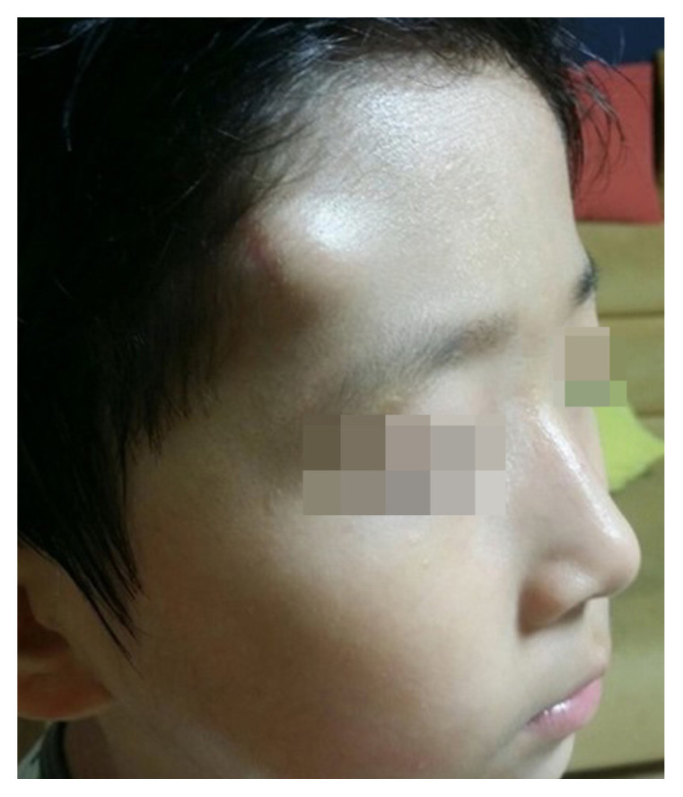
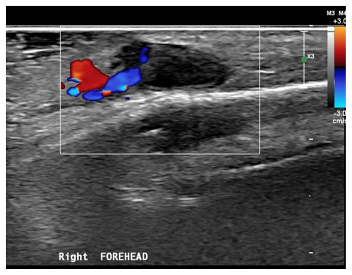
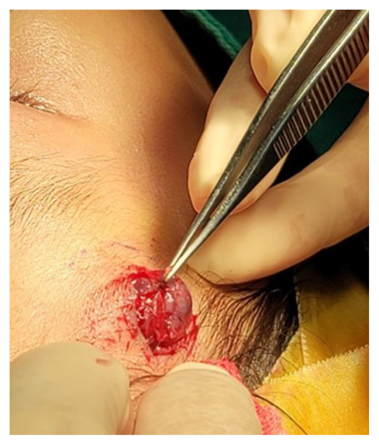
The superficial temporal artery (STA), the terminal branch of the external carotid artery, is divided into the frontal (anterior) and parietal (posterior) branches. The frontal branch of the STA is located superficially on the anterior region of the scalp, making it especially susceptible to trauma. Here, we report a traumatic pseudoaneurysm of the STA in a 7-year-old boy who was injured in a minor car accident. A physical examination showed only a small bruise on the patient's forehead, and all vital signs were stable at the emergency room of our medical center. A facial computed tomography scan showed no significant findings. However, the boy later re-visited the hospital with slight swelling on the right forehead, and an ultrasonography scan revealed a hematoma near the right temporal artery. The resected hematoma (approximately 2 cm) was diagnosed as a traumatic pseudoaneurysm. Awareness of the possibility of a traumatic pseudoaneurysm in the STA may prevent a circumspect diagnosis in the future.


 分享
分享
 求助内容:
求助内容: 应助结果提醒方式:
 扫码关注我们
