Kyril L Cole, Matthew C Findlay, Mrinmoy Kundu, Chase Johansen, Cameron Rawanduzy, Brandon Lucke-Wold
{"title":"先进成像技术在神经外科诊断中的作用。","authors":"Kyril L Cole, Matthew C Findlay, Mrinmoy Kundu, Chase Johansen, Cameron Rawanduzy, Brandon Lucke-Wold","doi":"10.53964/jmmi.2023002","DOIUrl":null,"url":null,"abstract":"<p><p>Neurosurgery as a specialty has developed at a rapid pace as a result of the continual advancements in neuroimaging modalities. With more sophisticated imaging options available to the modern neurosurgeon, diagnoses become more accurate and at a faster rate, allowing for greater surgical planning and precision. Herein, the authors review the current heavily used imaging modalities within neurosurgery, weighing their strengths and weaknesses, and provide a look into new advances and imaging options within the field. Of the many imaging modalities currently available to the practicing neurosurgeon, magnetic resonance imaging (MRI), computed tomography (CT), positron emission tomography (PET), and ultrasonography (US) are used most heavily within the field for appropriate diagnosis of neuropathologies in question. For each, their strengths are weighed regarding appropriate capabilities in accurate diagnosis of cranial or spinal lesions. Reasoning for choosing one over the other for various pathologies is also reviewed. Current limitations of each is also assessed, providing insight for possible improvement for each. New advancements in imaging options are subsequently reviewed for best uses within neurosurgery, including the new utilization of FIESTA sequencing, glymphatic mapping, black-blood MRI, and functional MRI. The specialty of neurosurgery will continue to heavily rely on improvements within imaging options available for improved diagnosis and greater surgical outcomes for the patients treated. The synthesis of techniques provided herein may provide meaningful guidance for neurosurgeons in effectively diagnosing neurological pathologies while also helping guide future efforts in neuroimaging developments.</p>","PeriodicalId":73833,"journal":{"name":"Journal of modern medical imaging","volume":"1 ","pages":""},"PeriodicalIF":0.0000,"publicationDate":"2023-01-01","publicationTypes":"Journal Article","fieldsOfStudy":null,"isOpenAccess":false,"openAccessPdf":"","citationCount":"0","resultStr":"{\"title\":\"The Role of Advanced Imaging in Neurosurgical Diagnosis.\",\"authors\":\"Kyril L Cole, Matthew C Findlay, Mrinmoy Kundu, Chase Johansen, Cameron Rawanduzy, Brandon Lucke-Wold\",\"doi\":\"10.53964/jmmi.2023002\",\"DOIUrl\":null,\"url\":null,\"abstract\":\"<p><p>Neurosurgery as a specialty has developed at a rapid pace as a result of the continual advancements in neuroimaging modalities. With more sophisticated imaging options available to the modern neurosurgeon, diagnoses become more accurate and at a faster rate, allowing for greater surgical planning and precision. Herein, the authors review the current heavily used imaging modalities within neurosurgery, weighing their strengths and weaknesses, and provide a look into new advances and imaging options within the field. Of the many imaging modalities currently available to the practicing neurosurgeon, magnetic resonance imaging (MRI), computed tomography (CT), positron emission tomography (PET), and ultrasonography (US) are used most heavily within the field for appropriate diagnosis of neuropathologies in question. For each, their strengths are weighed regarding appropriate capabilities in accurate diagnosis of cranial or spinal lesions. Reasoning for choosing one over the other for various pathologies is also reviewed. Current limitations of each is also assessed, providing insight for possible improvement for each. New advancements in imaging options are subsequently reviewed for best uses within neurosurgery, including the new utilization of FIESTA sequencing, glymphatic mapping, black-blood MRI, and functional MRI. The specialty of neurosurgery will continue to heavily rely on improvements within imaging options available for improved diagnosis and greater surgical outcomes for the patients treated. The synthesis of techniques provided herein may provide meaningful guidance for neurosurgeons in effectively diagnosing neurological pathologies while also helping guide future efforts in neuroimaging developments.</p>\",\"PeriodicalId\":73833,\"journal\":{\"name\":\"Journal of modern medical imaging\",\"volume\":\"1 \",\"pages\":\"\"},\"PeriodicalIF\":0.0000,\"publicationDate\":\"2023-01-01\",\"publicationTypes\":\"Journal Article\",\"fieldsOfStudy\":null,\"isOpenAccess\":false,\"openAccessPdf\":\"\",\"citationCount\":\"0\",\"resultStr\":null,\"platform\":\"Semanticscholar\",\"paperid\":null,\"PeriodicalName\":\"Journal of modern medical imaging\",\"FirstCategoryId\":\"1085\",\"ListUrlMain\":\"https://doi.org/10.53964/jmmi.2023002\",\"RegionNum\":0,\"RegionCategory\":null,\"ArticlePicture\":[],\"TitleCN\":null,\"AbstractTextCN\":null,\"PMCID\":null,\"EPubDate\":\"2023/2/15 0:00:00\",\"PubModel\":\"Epub\",\"JCR\":\"\",\"JCRName\":\"\",\"Score\":null,\"Total\":0}","platform":"Semanticscholar","paperid":null,"PeriodicalName":"Journal of modern medical imaging","FirstCategoryId":"1085","ListUrlMain":"https://doi.org/10.53964/jmmi.2023002","RegionNum":0,"RegionCategory":null,"ArticlePicture":[],"TitleCN":null,"AbstractTextCN":null,"PMCID":null,"EPubDate":"2023/2/15 0:00:00","PubModel":"Epub","JCR":"","JCRName":"","Score":null,"Total":0}
The Role of Advanced Imaging in Neurosurgical Diagnosis.
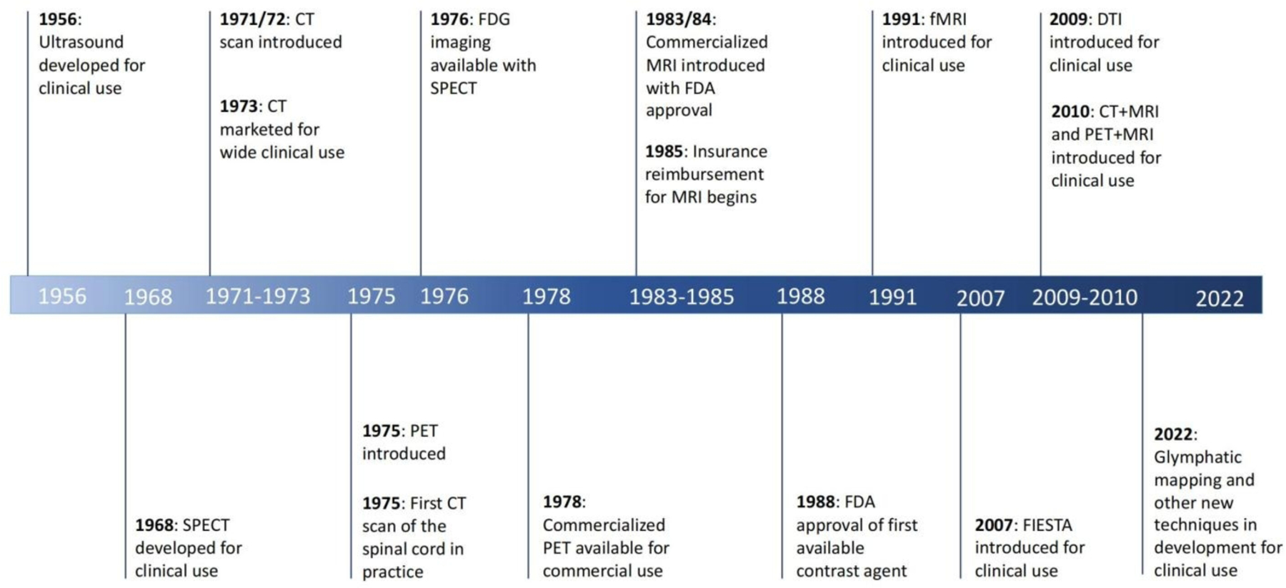
Neurosurgery as a specialty has developed at a rapid pace as a result of the continual advancements in neuroimaging modalities. With more sophisticated imaging options available to the modern neurosurgeon, diagnoses become more accurate and at a faster rate, allowing for greater surgical planning and precision. Herein, the authors review the current heavily used imaging modalities within neurosurgery, weighing their strengths and weaknesses, and provide a look into new advances and imaging options within the field. Of the many imaging modalities currently available to the practicing neurosurgeon, magnetic resonance imaging (MRI), computed tomography (CT), positron emission tomography (PET), and ultrasonography (US) are used most heavily within the field for appropriate diagnosis of neuropathologies in question. For each, their strengths are weighed regarding appropriate capabilities in accurate diagnosis of cranial or spinal lesions. Reasoning for choosing one over the other for various pathologies is also reviewed. Current limitations of each is also assessed, providing insight for possible improvement for each. New advancements in imaging options are subsequently reviewed for best uses within neurosurgery, including the new utilization of FIESTA sequencing, glymphatic mapping, black-blood MRI, and functional MRI. The specialty of neurosurgery will continue to heavily rely on improvements within imaging options available for improved diagnosis and greater surgical outcomes for the patients treated. The synthesis of techniques provided herein may provide meaningful guidance for neurosurgeons in effectively diagnosing neurological pathologies while also helping guide future efforts in neuroimaging developments.


 分享
分享

 求助内容:
求助内容: 应助结果提醒方式:
 扫码关注我们
