Yong Jig Lee, Dong Gil Han, Se Hun Kim, Jeong Su Shim, Sung-Eun Kim
{"title":"孤立性颧弓骨折软组织与骨凹陷位置的差异。","authors":"Yong Jig Lee, Dong Gil Han, Se Hun Kim, Jeong Su Shim, Sung-Eun Kim","doi":"10.7181/acfs.2023.00031","DOIUrl":null,"url":null,"abstract":"<p><strong>Background: </strong>When performing reduction of zygomatic arch fractures, locating the inward portion of the fracture can be difficult. Therefore, this study investigated the discrepancy between the locations of the depression on the soft tissue and bone and sought to identify how to determine the inward portion of the fracture on the patient's face.</p><p><strong>Methods: </strong>We conducted a retrospective review of chart with isolated zygomatic arch fractures of type V in the Nam and Jung classification from March 2013 to February 2022. For consistent measurements, a reference point (RP), at the intersection between a vertical line passing through the end point of the root of the ear helix in the patient's side-view photograph and a transverse line passing through the longest horizontal axis of the external meatus opening, was established. We then measured the distance between the RP and the soft tissue depression in a portrait and the bone depression on a computed tomography (CT) scan. The discrepancy between these distances was quantified.</p><p><strong>Results: </strong>Among the patients with isolated zygomatic arch fractures, only those with a fully visible ear on a side-view photograph were included. Twenty-four patients met the inclusion criteria. There were four types of discrepancies in the location of the soft tissue depression compared to the bone depression: type I, forward and upward discrepancy (7.45 and 3.28 mm), type II, backward and upward (4.29 and 4.21 mm), type III, forward and downward (10.06 and 5.15 mm), and type IV, backward and downward (2.61 and 3.27 mm).</p><p><strong>Conclusion: </strong>This study showed that discrepancy between the locations of the depressions on the soft tissue and bone exists in various directions. Therefore, applying the transverse and vertical distances measured from a bone image of the CT scan onto the patient's face at the indicated RP will be helpful for predicting the reduction location.</p>","PeriodicalId":52238,"journal":{"name":"Archives of Craniofacial Surgery","volume":"24 1","pages":"18-23"},"PeriodicalIF":0.0000,"publicationDate":"2023-02-01","publicationTypes":"Journal Article","fieldsOfStudy":null,"isOpenAccess":false,"openAccessPdf":"https://ftp.ncbi.nlm.nih.gov/pub/pmc/oa_pdf/86/dc/acfs-2023-00031.PMC10009208.pdf","citationCount":"0","resultStr":"{\"title\":\"Discrepancy of the location of depression on the soft tissue and the bone in isolated zygomatic arch fracture.\",\"authors\":\"Yong Jig Lee, Dong Gil Han, Se Hun Kim, Jeong Su Shim, Sung-Eun Kim\",\"doi\":\"10.7181/acfs.2023.00031\",\"DOIUrl\":null,\"url\":null,\"abstract\":\"<p><strong>Background: </strong>When performing reduction of zygomatic arch fractures, locating the inward portion of the fracture can be difficult. Therefore, this study investigated the discrepancy between the locations of the depression on the soft tissue and bone and sought to identify how to determine the inward portion of the fracture on the patient's face.</p><p><strong>Methods: </strong>We conducted a retrospective review of chart with isolated zygomatic arch fractures of type V in the Nam and Jung classification from March 2013 to February 2022. For consistent measurements, a reference point (RP), at the intersection between a vertical line passing through the end point of the root of the ear helix in the patient's side-view photograph and a transverse line passing through the longest horizontal axis of the external meatus opening, was established. We then measured the distance between the RP and the soft tissue depression in a portrait and the bone depression on a computed tomography (CT) scan. The discrepancy between these distances was quantified.</p><p><strong>Results: </strong>Among the patients with isolated zygomatic arch fractures, only those with a fully visible ear on a side-view photograph were included. Twenty-four patients met the inclusion criteria. There were four types of discrepancies in the location of the soft tissue depression compared to the bone depression: type I, forward and upward discrepancy (7.45 and 3.28 mm), type II, backward and upward (4.29 and 4.21 mm), type III, forward and downward (10.06 and 5.15 mm), and type IV, backward and downward (2.61 and 3.27 mm).</p><p><strong>Conclusion: </strong>This study showed that discrepancy between the locations of the depressions on the soft tissue and bone exists in various directions. Therefore, applying the transverse and vertical distances measured from a bone image of the CT scan onto the patient's face at the indicated RP will be helpful for predicting the reduction location.</p>\",\"PeriodicalId\":52238,\"journal\":{\"name\":\"Archives of Craniofacial Surgery\",\"volume\":\"24 1\",\"pages\":\"18-23\"},\"PeriodicalIF\":0.0000,\"publicationDate\":\"2023-02-01\",\"publicationTypes\":\"Journal Article\",\"fieldsOfStudy\":null,\"isOpenAccess\":false,\"openAccessPdf\":\"https://ftp.ncbi.nlm.nih.gov/pub/pmc/oa_pdf/86/dc/acfs-2023-00031.PMC10009208.pdf\",\"citationCount\":\"0\",\"resultStr\":null,\"platform\":\"Semanticscholar\",\"paperid\":null,\"PeriodicalName\":\"Archives of Craniofacial Surgery\",\"FirstCategoryId\":\"1085\",\"ListUrlMain\":\"https://doi.org/10.7181/acfs.2023.00031\",\"RegionNum\":0,\"RegionCategory\":null,\"ArticlePicture\":[],\"TitleCN\":null,\"AbstractTextCN\":null,\"PMCID\":null,\"EPubDate\":\"\",\"PubModel\":\"\",\"JCR\":\"Q2\",\"JCRName\":\"Medicine\",\"Score\":null,\"Total\":0}","platform":"Semanticscholar","paperid":null,"PeriodicalName":"Archives of Craniofacial Surgery","FirstCategoryId":"1085","ListUrlMain":"https://doi.org/10.7181/acfs.2023.00031","RegionNum":0,"RegionCategory":null,"ArticlePicture":[],"TitleCN":null,"AbstractTextCN":null,"PMCID":null,"EPubDate":"","PubModel":"","JCR":"Q2","JCRName":"Medicine","Score":null,"Total":0}
Discrepancy of the location of depression on the soft tissue and the bone in isolated zygomatic arch fracture.
Background: When performing reduction of zygomatic arch fractures, locating the inward portion of the fracture can be difficult. Therefore, this study investigated the discrepancy between the locations of the depression on the soft tissue and bone and sought to identify how to determine the inward portion of the fracture on the patient's face.
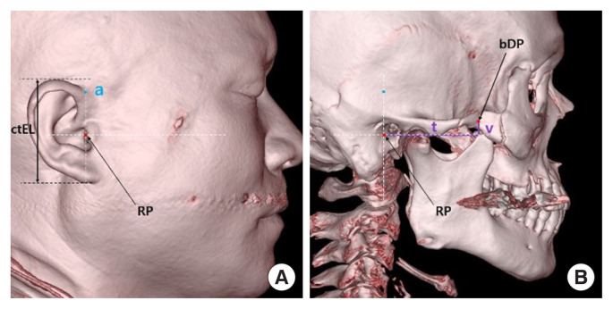
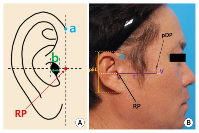
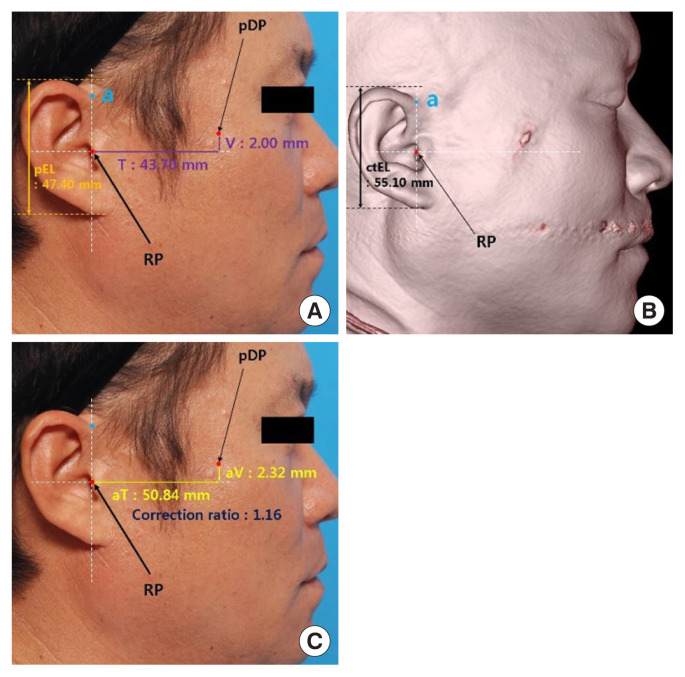
Methods: We conducted a retrospective review of chart with isolated zygomatic arch fractures of type V in the Nam and Jung classification from March 2013 to February 2022. For consistent measurements, a reference point (RP), at the intersection between a vertical line passing through the end point of the root of the ear helix in the patient's side-view photograph and a transverse line passing through the longest horizontal axis of the external meatus opening, was established. We then measured the distance between the RP and the soft tissue depression in a portrait and the bone depression on a computed tomography (CT) scan. The discrepancy between these distances was quantified.
Results: Among the patients with isolated zygomatic arch fractures, only those with a fully visible ear on a side-view photograph were included. Twenty-four patients met the inclusion criteria. There were four types of discrepancies in the location of the soft tissue depression compared to the bone depression: type I, forward and upward discrepancy (7.45 and 3.28 mm), type II, backward and upward (4.29 and 4.21 mm), type III, forward and downward (10.06 and 5.15 mm), and type IV, backward and downward (2.61 and 3.27 mm).
Conclusion: This study showed that discrepancy between the locations of the depressions on the soft tissue and bone exists in various directions. Therefore, applying the transverse and vertical distances measured from a bone image of the CT scan onto the patient's face at the indicated RP will be helpful for predicting the reduction location.


 分享
分享
 求助内容:
求助内容: 应助结果提醒方式:
 扫码关注我们
