Edmundo J Nassri Camara, Flávia R do Prado Valladares, Ng Kin Key, Paloma Fonseca Santana, Jun Ramos Kawaoka, Thais Harada Campos, Marcus Ribeiro de O Santana, Alex Costa Cunha, Danilo Sousa Sampaio, Gustavo Pinheiro Santana, Luis Gustavo S Brito, Narjara de O Cardoso Dourado, Saulo Jende Nascimento, Alice Povoa A L Lira, Naily N do Nascimento, Romeu Pacheco F Dos Santos, Sérgio Rodrigo F Rocha, Thaise Gordiano Machado
{"title":"超声心动图显示巴西无心脏病和心脏正常人群的体表面积和身高对左心房容量的指标性影响肥胖和超重患者的行为。","authors":"Edmundo J Nassri Camara, Flávia R do Prado Valladares, Ng Kin Key, Paloma Fonseca Santana, Jun Ramos Kawaoka, Thais Harada Campos, Marcus Ribeiro de O Santana, Alex Costa Cunha, Danilo Sousa Sampaio, Gustavo Pinheiro Santana, Luis Gustavo S Brito, Narjara de O Cardoso Dourado, Saulo Jende Nascimento, Alice Povoa A L Lira, Naily N do Nascimento, Romeu Pacheco F Dos Santos, Sérgio Rodrigo F Rocha, Thaise Gordiano Machado","doi":"10.26502/fccm.92920304","DOIUrl":null,"url":null,"abstract":"<p><strong>Background: </strong>Left atrial (LA) volume indexing for body surface area (BSA) may underestimate LA size in obese and overweight people. Since LA volume is a risk marker for some cardiovascular events, it is suggested that indexing for height would be an alternative more appropriate method. The aims of this study were to find normal and the best cutoff values for LA volume indexed for height in our population.</p><p><strong>Methods: </strong>Echocardiograms from 2018 to 2021 were reviewed and patients without known cardiac disease and completely normal echocardiograms that had the left atrial volume (LAvol) measured by biplane Simpson's method were included. LAvol was indexed by BSA (ml/m<sup>2</sup>), by height (LAvol/m), by height raised to exponent 2.7 (ml/ m<sup>2.7</sup>) and by height squared (ml/h<sup>2</sup>).</p><p><strong>Results: </strong>A total of 545 patients, 50.5 ± 13.4 y., 335 females (61,5%) were analyzed. There were 145 normal weight (26.6%), 215 overweight (39.4%), 154 obese (28.3%) and 31 low weight (5.7%) patients. To establish normal values we included only the normal weight group and considered normal values from 2SD below to 2SD above the mean. Mean and normal values were: LAvol/h 26.0 ±4.5, 17 - 35 ml/m, LAvol/ht<sup>2</sup> 16 ± 2.8, 10.4 - 21.6 ml/ ht<sup>2</sup> and LAvol/ht<sup>2.7</sup> 11.4 ± 2.2, 7.0 - 15.8 ml/m<sup>2.7</sup>. The normal LAvol/ht<sup>2.7</sup> differed between male and female (11.4 ± 2.4 and 12.8 ± 2.6, p < 0.001). LA diameter, LAvol, LAvol/h, LAvol/h<sup>2</sup> and LAvol/ht<sup>2.7</sup> increased progressively from low-weight, normal weight, overweight and obese patients (p< 0.0001), but not LAvol/BSA. When indexing LAvol for height, for height<sup>2</sup> and for height<sup>2.7</sup> 20.8%, 22.7% and 21.4% of the obese patients, respectively, were reclassified as enlarged LA, and 7.4%, 8.8% and 8.4% of the overweight patients as well. Using ROC curve analysis, LAvol/h<sup>2</sup> had the highest AUC ant the best predictive value to identify LA enlargement and LAvol/BSA the worst one.</p><p><strong>Conclusions: </strong>Normal values for LAvol indexed for height by three different methods are described in normal individuals. We reinforce that LAvol indexation for BSA underestimates LA size in obese and overweight patients and in these groups, specially, indexing for height<sup>2</sup> is probably the best method to evaluate LAvol.</p>","PeriodicalId":72523,"journal":{"name":"Cardiology and cardiovascular medicine","volume":"7 1","pages":"25-31"},"PeriodicalIF":0.0000,"publicationDate":"2023-01-01","publicationTypes":"Journal Article","fieldsOfStudy":null,"isOpenAccess":false,"openAccessPdf":"https://www.ncbi.nlm.nih.gov/pmc/articles/PMC10019596/pdf/","citationCount":"3","resultStr":"{\"title\":\"Indexing of Left Atrial Volume by Body Surface Area and Height in a Brazilian Population without Previous Heart Disease and with a Normal Heart on Echocardiography. Behavior in Obese and Overweight Patients.\",\"authors\":\"Edmundo J Nassri Camara, Flávia R do Prado Valladares, Ng Kin Key, Paloma Fonseca Santana, Jun Ramos Kawaoka, Thais Harada Campos, Marcus Ribeiro de O Santana, Alex Costa Cunha, Danilo Sousa Sampaio, Gustavo Pinheiro Santana, Luis Gustavo S Brito, Narjara de O Cardoso Dourado, Saulo Jende Nascimento, Alice Povoa A L Lira, Naily N do Nascimento, Romeu Pacheco F Dos Santos, Sérgio Rodrigo F Rocha, Thaise Gordiano Machado\",\"doi\":\"10.26502/fccm.92920304\",\"DOIUrl\":null,\"url\":null,\"abstract\":\"<p><strong>Background: </strong>Left atrial (LA) volume indexing for body surface area (BSA) may underestimate LA size in obese and overweight people. Since LA volume is a risk marker for some cardiovascular events, it is suggested that indexing for height would be an alternative more appropriate method. The aims of this study were to find normal and the best cutoff values for LA volume indexed for height in our population.</p><p><strong>Methods: </strong>Echocardiograms from 2018 to 2021 were reviewed and patients without known cardiac disease and completely normal echocardiograms that had the left atrial volume (LAvol) measured by biplane Simpson's method were included. LAvol was indexed by BSA (ml/m<sup>2</sup>), by height (LAvol/m), by height raised to exponent 2.7 (ml/ m<sup>2.7</sup>) and by height squared (ml/h<sup>2</sup>).</p><p><strong>Results: </strong>A total of 545 patients, 50.5 ± 13.4 y., 335 females (61,5%) were analyzed. There were 145 normal weight (26.6%), 215 overweight (39.4%), 154 obese (28.3%) and 31 low weight (5.7%) patients. To establish normal values we included only the normal weight group and considered normal values from 2SD below to 2SD above the mean. Mean and normal values were: LAvol/h 26.0 ±4.5, 17 - 35 ml/m, LAvol/ht<sup>2</sup> 16 ± 2.8, 10.4 - 21.6 ml/ ht<sup>2</sup> and LAvol/ht<sup>2.7</sup> 11.4 ± 2.2, 7.0 - 15.8 ml/m<sup>2.7</sup>. The normal LAvol/ht<sup>2.7</sup> differed between male and female (11.4 ± 2.4 and 12.8 ± 2.6, p < 0.001). LA diameter, LAvol, LAvol/h, LAvol/h<sup>2</sup> and LAvol/ht<sup>2.7</sup> increased progressively from low-weight, normal weight, overweight and obese patients (p< 0.0001), but not LAvol/BSA. When indexing LAvol for height, for height<sup>2</sup> and for height<sup>2.7</sup> 20.8%, 22.7% and 21.4% of the obese patients, respectively, were reclassified as enlarged LA, and 7.4%, 8.8% and 8.4% of the overweight patients as well. Using ROC curve analysis, LAvol/h<sup>2</sup> had the highest AUC ant the best predictive value to identify LA enlargement and LAvol/BSA the worst one.</p><p><strong>Conclusions: </strong>Normal values for LAvol indexed for height by three different methods are described in normal individuals. We reinforce that LAvol indexation for BSA underestimates LA size in obese and overweight patients and in these groups, specially, indexing for height<sup>2</sup> is probably the best method to evaluate LAvol.</p>\",\"PeriodicalId\":72523,\"journal\":{\"name\":\"Cardiology and cardiovascular medicine\",\"volume\":\"7 1\",\"pages\":\"25-31\"},\"PeriodicalIF\":0.0000,\"publicationDate\":\"2023-01-01\",\"publicationTypes\":\"Journal Article\",\"fieldsOfStudy\":null,\"isOpenAccess\":false,\"openAccessPdf\":\"https://www.ncbi.nlm.nih.gov/pmc/articles/PMC10019596/pdf/\",\"citationCount\":\"3\",\"resultStr\":null,\"platform\":\"Semanticscholar\",\"paperid\":null,\"PeriodicalName\":\"Cardiology and cardiovascular medicine\",\"FirstCategoryId\":\"1085\",\"ListUrlMain\":\"https://doi.org/10.26502/fccm.92920304\",\"RegionNum\":0,\"RegionCategory\":null,\"ArticlePicture\":[],\"TitleCN\":null,\"AbstractTextCN\":null,\"PMCID\":null,\"EPubDate\":\"\",\"PubModel\":\"\",\"JCR\":\"\",\"JCRName\":\"\",\"Score\":null,\"Total\":0}","platform":"Semanticscholar","paperid":null,"PeriodicalName":"Cardiology and cardiovascular medicine","FirstCategoryId":"1085","ListUrlMain":"https://doi.org/10.26502/fccm.92920304","RegionNum":0,"RegionCategory":null,"ArticlePicture":[],"TitleCN":null,"AbstractTextCN":null,"PMCID":null,"EPubDate":"","PubModel":"","JCR":"","JCRName":"","Score":null,"Total":0}
Indexing of Left Atrial Volume by Body Surface Area and Height in a Brazilian Population without Previous Heart Disease and with a Normal Heart on Echocardiography. Behavior in Obese and Overweight Patients.
Background: Left atrial (LA) volume indexing for body surface area (BSA) may underestimate LA size in obese and overweight people. Since LA volume is a risk marker for some cardiovascular events, it is suggested that indexing for height would be an alternative more appropriate method. The aims of this study were to find normal and the best cutoff values for LA volume indexed for height in our population.
Methods: Echocardiograms from 2018 to 2021 were reviewed and patients without known cardiac disease and completely normal echocardiograms that had the left atrial volume (LAvol) measured by biplane Simpson's method were included. LAvol was indexed by BSA (ml/m2), by height (LAvol/m), by height raised to exponent 2.7 (ml/ m2.7) and by height squared (ml/h2).
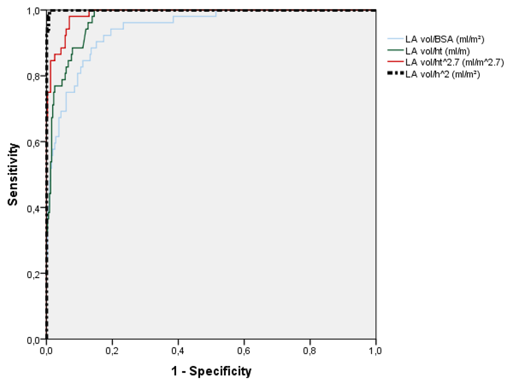
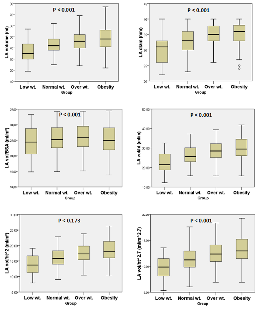
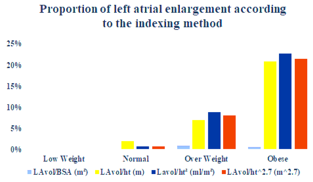
Results: A total of 545 patients, 50.5 ± 13.4 y., 335 females (61,5%) were analyzed. There were 145 normal weight (26.6%), 215 overweight (39.4%), 154 obese (28.3%) and 31 low weight (5.7%) patients. To establish normal values we included only the normal weight group and considered normal values from 2SD below to 2SD above the mean. Mean and normal values were: LAvol/h 26.0 ±4.5, 17 - 35 ml/m, LAvol/ht2 16 ± 2.8, 10.4 - 21.6 ml/ ht2 and LAvol/ht2.7 11.4 ± 2.2, 7.0 - 15.8 ml/m2.7. The normal LAvol/ht2.7 differed between male and female (11.4 ± 2.4 and 12.8 ± 2.6, p < 0.001). LA diameter, LAvol, LAvol/h, LAvol/h2 and LAvol/ht2.7 increased progressively from low-weight, normal weight, overweight and obese patients (p< 0.0001), but not LAvol/BSA. When indexing LAvol for height, for height2 and for height2.7 20.8%, 22.7% and 21.4% of the obese patients, respectively, were reclassified as enlarged LA, and 7.4%, 8.8% and 8.4% of the overweight patients as well. Using ROC curve analysis, LAvol/h2 had the highest AUC ant the best predictive value to identify LA enlargement and LAvol/BSA the worst one.
Conclusions: Normal values for LAvol indexed for height by three different methods are described in normal individuals. We reinforce that LAvol indexation for BSA underestimates LA size in obese and overweight patients and in these groups, specially, indexing for height2 is probably the best method to evaluate LAvol.


 分享
分享

 求助内容:
求助内容: 应助结果提醒方式:
 扫码关注我们
